Stefano Piscone, Sara Saccone, Paola Milillo, Giorgia Schiraldi, Roberta Vinci, Luca Macarini, Luca Pio Stoppino
{"title":"Bone Mineral Density (BMD) Assessment Using Dual-Energy CT with Different Base Material Pairs (BMPs).","authors":"Stefano Piscone, Sara Saccone, Paola Milillo, Giorgia Schiraldi, Roberta Vinci, Luca Macarini, Luca Pio Stoppino","doi":"10.3390/jimaging11070236","DOIUrl":null,"url":null,"abstract":"<p><p>The assessment of bone mineral density (BMD) is essential for osteoporosis diagnosis. Dual-energy X-ray Absorptiometry (DXA) is the current gold standard, but it has limitations in evaluating trabecular bone and is susceptible to different artifacts. In this study we evaluate whether Dual-Energy Computed Tomography (DECT) can be defined as an alternative method for the assessment of BMD in a sample of postmenopausal patients undergoing oncological follow-up. In this study a retrospective analysis was conducted on 41 patients who had both DECT and DXA within six months. BMD values were extracted from DECT using five different base material pairs (BMPs) and compared with DXA measurements at the femoral neck. The calcium-fat pairing showed the strongest correlation with DXA-derived BMD (Spearman's ρ = 0.797) and excellent reproducibility (ICC = 0.983). There was a strong and significant association between the DXA results and the various BPM measurements. These findings support the possibility of DECT in the precise and opportunistic evaluation of BMD changes when employing particular BMPs. This study showed how this technique can be a useful and effective substitute for conventional DXA, particularly when patients are in oncological follow-up using DECT, minimizing additional radiation exposure.</p>","PeriodicalId":37035,"journal":{"name":"Journal of Imaging","volume":"11 7","pages":""},"PeriodicalIF":2.7000,"publicationDate":"2025-07-13","publicationTypes":"Journal Article","fieldsOfStudy":null,"isOpenAccess":false,"openAccessPdf":"https://www.ncbi.nlm.nih.gov/pmc/articles/PMC12295864/pdf/","citationCount":"0","resultStr":null,"platform":"Semanticscholar","paperid":null,"PeriodicalName":"Journal of Imaging","FirstCategoryId":"1085","ListUrlMain":"https://doi.org/10.3390/jimaging11070236","RegionNum":0,"RegionCategory":null,"ArticlePicture":[],"TitleCN":null,"AbstractTextCN":null,"PMCID":null,"EPubDate":"","PubModel":"","JCR":"Q3","JCRName":"IMAGING SCIENCE & PHOTOGRAPHIC TECHNOLOGY","Score":null,"Total":0}
引用次数: 0
Abstract
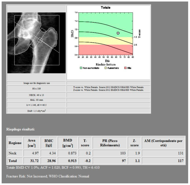
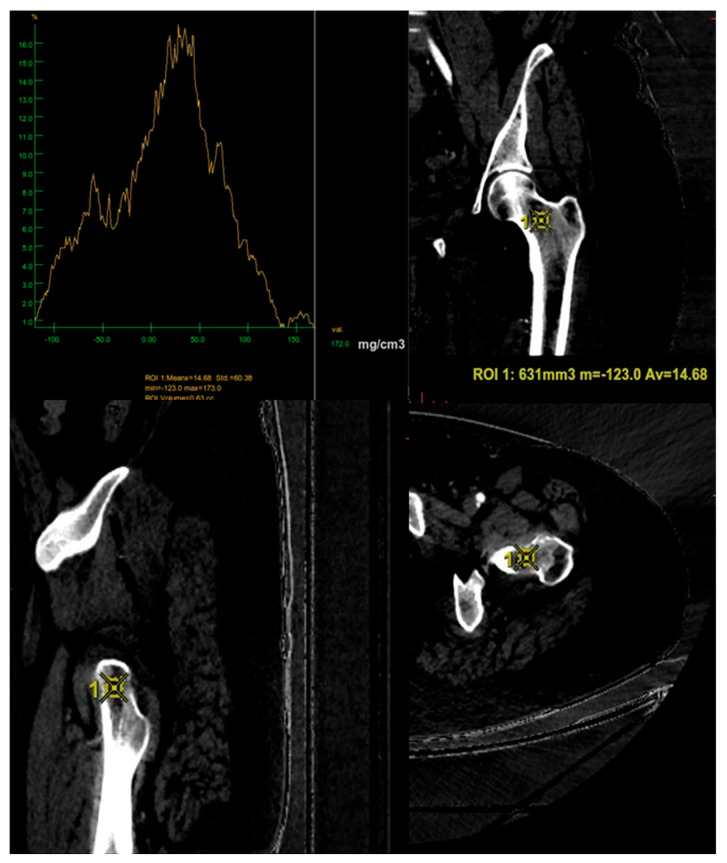
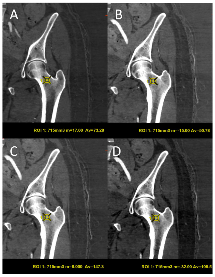
The assessment of bone mineral density (BMD) is essential for osteoporosis diagnosis. Dual-energy X-ray Absorptiometry (DXA) is the current gold standard, but it has limitations in evaluating trabecular bone and is susceptible to different artifacts. In this study we evaluate whether Dual-Energy Computed Tomography (DECT) can be defined as an alternative method for the assessment of BMD in a sample of postmenopausal patients undergoing oncological follow-up. In this study a retrospective analysis was conducted on 41 patients who had both DECT and DXA within six months. BMD values were extracted from DECT using five different base material pairs (BMPs) and compared with DXA measurements at the femoral neck. The calcium-fat pairing showed the strongest correlation with DXA-derived BMD (Spearman's ρ = 0.797) and excellent reproducibility (ICC = 0.983). There was a strong and significant association between the DXA results and the various BPM measurements. These findings support the possibility of DECT in the precise and opportunistic evaluation of BMD changes when employing particular BMPs. This study showed how this technique can be a useful and effective substitute for conventional DXA, particularly when patients are in oncological follow-up using DECT, minimizing additional radiation exposure.

 求助内容:
求助内容: 应助结果提醒方式:


