Najla Sadat, John Habakuk Lojenburg, Michael Scharfschwerdt, Matthias Klinger, Stephan Ensminger
{"title":"New Reproducible Porcine Aortic Root Calcification Model: An Ex Vivo Study Under Dynamic Conditions","authors":"Najla Sadat, John Habakuk Lojenburg, Michael Scharfschwerdt, Matthias Klinger, Stephan Ensminger","doi":"10.1155/jocs/5519548","DOIUrl":null,"url":null,"abstract":"<div>\n <p><b>Background:</b> Calcific aortic valve disease results in severe aortic valve stenosis, the most frequent valvular disorder. Ex vivo animal models provide an essential resource to understand the mechanism of valvular calcification. Therefore, we aimed to develop a new ex vivo porcine aortic root calcification model.</p>\n <p><b>Methods:</b> Porcine aortic roots were subjected to a procalcific treatment with a buffer solution containing a defined calcium concentration (CaCl<sub>2</sub> = 2.2 mmol) in a durability tester (Hi-Cycle tester) with over 53.2 million cycles. The control group consisted of native porcine aortic roots that were not treated. Cusps of porcine aortic valves of both groups (total <i>n</i> = 10) were compared through macroscopic evaluation, analysis of tissue thickness, scanning and transmission electron microscopy, histological examination and calcium determination.</p>\n <p><b>Results:</b> After durability testing, macroscopic examination demonstrated pronounced calcification at regions with high mechanical stress—the commissures and the nadirs of the cusps. Calcific nodules cause tissue thickness after Hi-Cycle testing. Hydroxyapatite crystals were found by scanning electron microscopy, and calcium deposits were noticed by transmission electron microscopy within calcified cusps in the calcified group. The proof of cusp calcification was seen histologically in the calcified group. Calcium content of the aortic cusps differed significantly after treatment with calcification buffer vs control group (7.240 [6.383–9.494] vs. 3.178 [3.140–3.701] μg/cm<sup>2</sup> cusp area, <i>p</i> = 0.008).</p>\n <p><b>Conclusion:</b> We established a new reproducible and dynamic porcine aortic root calcification model. This ex vivo model may be a helpful alternative for investigating treatment modalities of calcification and functional analysis of heart valves instead of a complex animal model.</p>\n </div>","PeriodicalId":15367,"journal":{"name":"Journal of Cardiac Surgery","volume":"2025 1","pages":""},"PeriodicalIF":1.3000,"publicationDate":"2025-07-26","publicationTypes":"Journal Article","fieldsOfStudy":null,"isOpenAccess":false,"openAccessPdf":"https://onlinelibrary.wiley.com/doi/epdf/10.1155/jocs/5519548","citationCount":"0","resultStr":null,"platform":"Semanticscholar","paperid":null,"PeriodicalName":"Journal of Cardiac Surgery","FirstCategoryId":"3","ListUrlMain":"https://onlinelibrary.wiley.com/doi/10.1155/jocs/5519548","RegionNum":4,"RegionCategory":"医学","ArticlePicture":[],"TitleCN":null,"AbstractTextCN":null,"PMCID":null,"EPubDate":"","PubModel":"","JCR":"Q3","JCRName":"CARDIAC & CARDIOVASCULAR SYSTEMS","Score":null,"Total":0}
引用次数: 0
Abstract
Background: Calcific aortic valve disease results in severe aortic valve stenosis, the most frequent valvular disorder. Ex vivo animal models provide an essential resource to understand the mechanism of valvular calcification. Therefore, we aimed to develop a new ex vivo porcine aortic root calcification model.
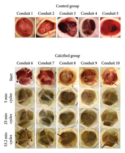
Methods: Porcine aortic roots were subjected to a procalcific treatment with a buffer solution containing a defined calcium concentration (CaCl2 = 2.2 mmol) in a durability tester (Hi-Cycle tester) with over 53.2 million cycles. The control group consisted of native porcine aortic roots that were not treated. Cusps of porcine aortic valves of both groups (total n = 10) were compared through macroscopic evaluation, analysis of tissue thickness, scanning and transmission electron microscopy, histological examination and calcium determination.
Results: After durability testing, macroscopic examination demonstrated pronounced calcification at regions with high mechanical stress—the commissures and the nadirs of the cusps. Calcific nodules cause tissue thickness after Hi-Cycle testing. Hydroxyapatite crystals were found by scanning electron microscopy, and calcium deposits were noticed by transmission electron microscopy within calcified cusps in the calcified group. The proof of cusp calcification was seen histologically in the calcified group. Calcium content of the aortic cusps differed significantly after treatment with calcification buffer vs control group (7.240 [6.383–9.494] vs. 3.178 [3.140–3.701] μg/cm2 cusp area, p = 0.008).
Conclusion: We established a new reproducible and dynamic porcine aortic root calcification model. This ex vivo model may be a helpful alternative for investigating treatment modalities of calcification and functional analysis of heart valves instead of a complex animal model.
期刊介绍:
Journal of Cardiac Surgery (JCS) is a peer-reviewed journal devoted to contemporary surgical treatment of cardiac disease. Renown for its detailed "how to" methods, JCS''s well-illustrated, concise technical articles, critical reviews and commentaries are highly valued by dedicated readers worldwide.
With Editor-in-Chief Harold Lazar, MD and an internationally prominent editorial board, JCS continues its 20-year history as an important professional resource. Editorial coverage includes biologic support, mechanical cardiac assist and/or replacement and surgical techniques, and features current material on topics such as OPCAB surgery, stented and stentless valves, endovascular stent placement, atrial fibrillation, transplantation, percutaneous valve repair/replacement, left ventricular restoration surgery, immunobiology, and bridges to transplant and recovery.
In addition, special sections (Images in Cardiac Surgery, Cardiac Regeneration) and historical reviews stimulate reader interest. The journal also routinely publishes proceedings of important international symposia in a timely manner.
 求助内容:
求助内容: 应助结果提醒方式:


