Shu-Xian Wu, Xiao-Long Liang, Qin-Qin Zhu, Wei Wang, Li Jiang, Huan-Huan Chen, Shui Tian, Ming Qi
{"title":"Altered hippocampal subfield volumes are associated with memory and executive function in patients with type 2 diabetes mellitus.","authors":"Shu-Xian Wu, Xiao-Long Liang, Qin-Qin Zhu, Wei Wang, Li Jiang, Huan-Huan Chen, Shui Tian, Ming Qi","doi":"10.4239/wjd.v16.i7.104424","DOIUrl":null,"url":null,"abstract":"<p><strong>Background: </strong>Increasing evidence has shown that hippocampal damage serves as a marker of early cognitive decline in patients with type 2 diabetes mellitus (T2DM); however, the association between hippocampal subregion volume changes and cognitive decline in different dimensions remains unclear.</p><p><strong>Aim: </strong>To investigate changes in hippocampal subregion volumes in patients with T2DM and their relationship with cognitive function impairment.</p><p><strong>Methods: </strong>Sixty patients with T2DM and 32 healthy controls were recruited. All participants underwent a 3.0 T magnetic resonance scan and a series of clinical assessments. Hippocampal subfield volumes were determined using FreeSurfer 7.4.1. A two-sample <i>t</i>-test was used to evaluate group differences. Partial correlation analysis was performed to assess the relationship between hippocampal subregion volumes and cognitive function. <sup>a</sup> <i>P</i> < 0.05 was considered statistically significant.</p><p><strong>Results: </strong>Compared with controls, the volume of right hippocampus-amygdala transition area (<i>t</i> = -3.053, <i>P</i> = 0.003) in patients with T2DM was significantly reduced, which was negatively correlated with the required time of the Trail Making Test (TMT)-A (<i>r</i> = -0.331, <i>P</i> = 0.028) and TMT-B (<i>r</i> = -0.402, <i>P</i> = 0.007) and positively correlated with the scores of Symbol Digit Modalities Test (<i>r</i> = 0.381, <i>P</i> = 0.011), Auditory Verbal Learning Test (AVLT)-N7 (<i>r</i> = 0.309, <i>P</i> = 0.041), and Digital Span Test (<i>r</i> = 0.300, <i>P</i> = 0.048). The volume of the right molecular layer (<i>t</i> = -2.998, <i>P</i> = 0.004) was also significantly reduced, which was positively associated with the scores of AVLT-N7 (<i>r</i> = 0.311, <i>P</i> = 0.045). In addition, the left hippocampal fissure volume (<i>t</i> = 3.617, <i>P</i> = 0.002) was significantly increased in patients with T2DM.</p><p><strong>Conclusion: </strong>Declines in cognitive performance, especially memory and executive function, are linked to changes in the volumes of the right hippocampus-amygdala transition area and right molecular layer in patients with T2DM.</p>","PeriodicalId":48607,"journal":{"name":"World Journal of Diabetes","volume":"16 7","pages":"104424"},"PeriodicalIF":4.6000,"publicationDate":"2025-07-15","publicationTypes":"Journal Article","fieldsOfStudy":null,"isOpenAccess":false,"openAccessPdf":"https://www.ncbi.nlm.nih.gov/pmc/articles/PMC12278078/pdf/","citationCount":"0","resultStr":null,"platform":"Semanticscholar","paperid":null,"PeriodicalName":"World Journal of Diabetes","FirstCategoryId":"3","ListUrlMain":"https://doi.org/10.4239/wjd.v16.i7.104424","RegionNum":3,"RegionCategory":"医学","ArticlePicture":[],"TitleCN":null,"AbstractTextCN":null,"PMCID":null,"EPubDate":"","PubModel":"","JCR":"Q1","JCRName":"ENDOCRINOLOGY & METABOLISM","Score":null,"Total":0}
引用次数: 0
Abstract
Background: Increasing evidence has shown that hippocampal damage serves as a marker of early cognitive decline in patients with type 2 diabetes mellitus (T2DM); however, the association between hippocampal subregion volume changes and cognitive decline in different dimensions remains unclear.
Aim: To investigate changes in hippocampal subregion volumes in patients with T2DM and their relationship with cognitive function impairment.
Methods: Sixty patients with T2DM and 32 healthy controls were recruited. All participants underwent a 3.0 T magnetic resonance scan and a series of clinical assessments. Hippocampal subfield volumes were determined using FreeSurfer 7.4.1. A two-sample t-test was used to evaluate group differences. Partial correlation analysis was performed to assess the relationship between hippocampal subregion volumes and cognitive function. aP < 0.05 was considered statistically significant.
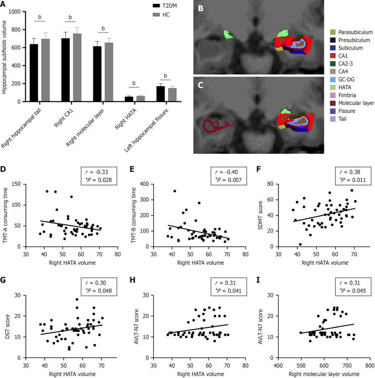
Results: Compared with controls, the volume of right hippocampus-amygdala transition area (t = -3.053, P = 0.003) in patients with T2DM was significantly reduced, which was negatively correlated with the required time of the Trail Making Test (TMT)-A (r = -0.331, P = 0.028) and TMT-B (r = -0.402, P = 0.007) and positively correlated with the scores of Symbol Digit Modalities Test (r = 0.381, P = 0.011), Auditory Verbal Learning Test (AVLT)-N7 (r = 0.309, P = 0.041), and Digital Span Test (r = 0.300, P = 0.048). The volume of the right molecular layer (t = -2.998, P = 0.004) was also significantly reduced, which was positively associated with the scores of AVLT-N7 (r = 0.311, P = 0.045). In addition, the left hippocampal fissure volume (t = 3.617, P = 0.002) was significantly increased in patients with T2DM.
Conclusion: Declines in cognitive performance, especially memory and executive function, are linked to changes in the volumes of the right hippocampus-amygdala transition area and right molecular layer in patients with T2DM.
期刊介绍:
The WJD is a high-quality, peer reviewed, open-access journal. The primary task of WJD is to rapidly publish high-quality original articles, reviews, editorials, and case reports in the field of diabetes. In order to promote productive academic communication, the peer review process for the WJD is transparent; to this end, all published manuscripts are accompanied by the anonymized reviewers’ comments as well as the authors’ responses. The primary aims of the WJD are to improve diagnostic, therapeutic and preventive modalities and the skills of clinicians and to guide clinical practice in diabetes. Scope: Diabetes Complications, Experimental Diabetes Mellitus, Type 1 Diabetes Mellitus, Type 2 Diabetes Mellitus, Diabetes, Gestational, Diabetic Angiopathies, Diabetic Cardiomyopathies, Diabetic Coma, Diabetic Ketoacidosis, Diabetic Nephropathies, Diabetic Neuropathies, Donohue Syndrome, Fetal Macrosomia, and Prediabetic State.
 求助内容:
求助内容: 应助结果提醒方式:


