Korbinian Krieger, Thomas Pyka, Clemens Mingels, Hasan Sari, Albert Gjedde, Axel Rominger, Paul Cumming
{"title":"Kinetics of the amino acid uptake tracer O-(2-[<sup>18</sup>F]fluoroethyl)-L-tyrosine (FET) in human brain.","authors":"Korbinian Krieger, Thomas Pyka, Clemens Mingels, Hasan Sari, Albert Gjedde, Axel Rominger, Paul Cumming","doi":"10.1186/s13550-025-01279-7","DOIUrl":null,"url":null,"abstract":"<p><strong>Background: </strong>The large neutral amino acid [<sup>18</sup>F]fluoroethoxy-L-tyrosine ([<sup>18</sup>F]FET) is a popular tracer for detection and staging of intracranial tumors by positron emission tomography (PET). While its high tumoral uptake reflects over-expression of the L-type amino acid transporter (LAT1), there is little knowledge about the kinetics of [<sup>18</sup>F]FET uptake in healthy brain tissue, owing to the limited PET data in healthy volunteers, and to the requirement of an arterial input function for compartmental analysis. To address this, we used long axial field-of-view (LAFOV) dynamic 40-min recordings of 28 post-operative patients with intracranial tumors to undertake parametric brain mapping relative to an image-derived arterial input function (IDIF) obtained from the aorta. We averaged the individual parametric maps to obtain estimates of the physiological uptake relatively unaffected by individual residual lesions and resections, and tested simplified single-frame methods for quantitation.</p><p><strong>Results: </strong>The analyses yielded estimates of regional unidirectional blood-brain clearance K<sub>1</sub> (0.00825-0.0244 ml g<sup>-1</sup> min<sup>-1</sup>), net blood-brain clearance K<sub>in</sub> (0.00448-0.00913 ml g<sup>-1</sup> min<sup>-1</sup>), and equilibrium distribution volume V<sub>T</sub> (0.126-0.495 ml g<sup>-1</sup>), where the lowest values depict white matter, and the highest values cerebellum. In our test of a simplified quantitation of [<sup>18</sup>F]FET uptake from single frame recordings, i.e., Gjedde-Patlak multilinear graphic analyses of K<sub>1</sub> at five min post-injection and K<sub>in</sub> at 40 min post injection, results were in good agreement with the analyses from the dynamic recordings (< 10% error).</p><p><strong>Conclusions: </strong>Compartmental analysis results for [<sup>18</sup>F]FET uptake in extra-tumoral human brain regions are in accord with the few prior reports, mainly obtained in experimental animals, and support the use of single frame quantitation. Present findings in relatively healthy brain should inform the interpretation of pathological [<sup>18</sup>F]FET uptake in tumors.</p>","PeriodicalId":11611,"journal":{"name":"EJNMMI Research","volume":"15 1","pages":"90"},"PeriodicalIF":3.1000,"publicationDate":"2025-07-22","publicationTypes":"Journal Article","fieldsOfStudy":null,"isOpenAccess":false,"openAccessPdf":"https://www.ncbi.nlm.nih.gov/pmc/articles/PMC12283531/pdf/","citationCount":"0","resultStr":null,"platform":"Semanticscholar","paperid":null,"PeriodicalName":"EJNMMI Research","FirstCategoryId":"3","ListUrlMain":"https://doi.org/10.1186/s13550-025-01279-7","RegionNum":3,"RegionCategory":"医学","ArticlePicture":[],"TitleCN":null,"AbstractTextCN":null,"PMCID":null,"EPubDate":"","PubModel":"","JCR":"Q1","JCRName":"RADIOLOGY, NUCLEAR MEDICINE & MEDICAL IMAGING","Score":null,"Total":0}
引用次数: 0
Abstract
Background: The large neutral amino acid [18F]fluoroethoxy-L-tyrosine ([18F]FET) is a popular tracer for detection and staging of intracranial tumors by positron emission tomography (PET). While its high tumoral uptake reflects over-expression of the L-type amino acid transporter (LAT1), there is little knowledge about the kinetics of [18F]FET uptake in healthy brain tissue, owing to the limited PET data in healthy volunteers, and to the requirement of an arterial input function for compartmental analysis. To address this, we used long axial field-of-view (LAFOV) dynamic 40-min recordings of 28 post-operative patients with intracranial tumors to undertake parametric brain mapping relative to an image-derived arterial input function (IDIF) obtained from the aorta. We averaged the individual parametric maps to obtain estimates of the physiological uptake relatively unaffected by individual residual lesions and resections, and tested simplified single-frame methods for quantitation.
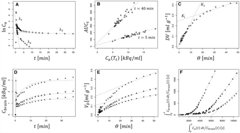
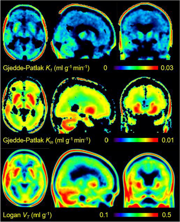
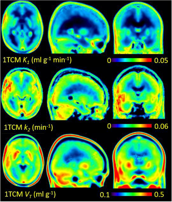
Results: The analyses yielded estimates of regional unidirectional blood-brain clearance K1 (0.00825-0.0244 ml g-1 min-1), net blood-brain clearance Kin (0.00448-0.00913 ml g-1 min-1), and equilibrium distribution volume VT (0.126-0.495 ml g-1), where the lowest values depict white matter, and the highest values cerebellum. In our test of a simplified quantitation of [18F]FET uptake from single frame recordings, i.e., Gjedde-Patlak multilinear graphic analyses of K1 at five min post-injection and Kin at 40 min post injection, results were in good agreement with the analyses from the dynamic recordings (< 10% error).
Conclusions: Compartmental analysis results for [18F]FET uptake in extra-tumoral human brain regions are in accord with the few prior reports, mainly obtained in experimental animals, and support the use of single frame quantitation. Present findings in relatively healthy brain should inform the interpretation of pathological [18F]FET uptake in tumors.
背景:大中性氨基酸[18F]氟乙氧基- l -酪氨酸([18F]FET)是一种常用的用于正电子发射断层扫描(PET)检测和分期颅内肿瘤的示踪剂。虽然FET在肿瘤中的高摄取反映了l型氨基酸转运蛋白(LAT1)的过度表达,但由于健康志愿者的PET数据有限,并且需要动脉输入功能进行区室分析,因此对[18F]FET在健康脑组织中的摄取动力学知之甚少。为了解决这个问题,我们使用长轴向视场(LAFOV)动态记录了28例术后颅内肿瘤患者的40分钟,并进行了相对于从主动脉获得的图像衍生动脉输入功能(IDIF)的参数化脑映射。我们取单个参数图的平均值,以获得相对不受单个残留病变和切除影响的生理摄取的估计值,并测试了简化的单帧定量方法。结果:分析得出了区域单向血脑清除率K1 (0.00825-0.0244 ml g-1 min-1),净血脑清除率Kin (0.00448-0.00913 ml g-1 min-1)和平衡分布体积VT (0.126-0.495 ml g-1)的估计值,其中最低值描绘白质,最高值描绘小脑。在我们对单帧记录的[18F]场效应管摄取的简化定量测试中,即对注射后5分钟的K1和注射后40分钟的Kin进行Gjedde-Patlak多线性图形分析,结果与动态记录的分析结果非常一致(结论:[18F]瘤外人脑区域FET摄取的区室分析结果与先前少数主要在实验动物中获得的报道一致,支持单帧定量的使用。目前在相对健康的大脑中的发现可以解释肿瘤中FET的病理性摄取[18F]。
EJNMMI ResearchRADIOLOGY, NUCLEAR MEDICINE & MEDICAL IMAGING&nb-
CiteScore
5.90
自引率
3.10%
发文量
72
审稿时长
13 weeks
期刊介绍:
EJNMMI Research publishes new basic, translational and clinical research in the field of nuclear medicine and molecular imaging. Regular features include original research articles, rapid communication of preliminary data on innovative research, interesting case reports, editorials, and letters to the editor. Educational articles on basic sciences, fundamental aspects and controversy related to pre-clinical and clinical research or ethical aspects of research are also welcome. Timely reviews provide updates on current applications, issues in imaging research and translational aspects of nuclear medicine and molecular imaging technologies.
The main emphasis is placed on the development of targeted imaging with radiopharmaceuticals within the broader context of molecular probes to enhance understanding and characterisation of the complex biological processes underlying disease and to develop, test and guide new treatment modalities, including radionuclide therapy.

 求助内容:
求助内容: 应助结果提醒方式:


