Shahram Oveisgharan, Armand Collin, Jingyun Yang, Sue E Leurgans, Veronique VanderHorst, David A Bennett, Julien Cohen-Adad, Osvaldo Delbono, Aron S Buchman
{"title":"Morphometric characteristics of tibial nerve and their relationship with age.","authors":"Shahram Oveisgharan, Armand Collin, Jingyun Yang, Sue E Leurgans, Veronique VanderHorst, David A Bennett, Julien Cohen-Adad, Osvaldo Delbono, Aron S Buchman","doi":"10.1093/braincomms/fcaf267","DOIUrl":null,"url":null,"abstract":"<p><p>Peripheral nerve comprises a crucial component of the distributed motor/sensory system. However, there is a paucity of data on peripheral nerve morphology derived from large numbers of older adults. This study aimed to quantify the morphometric characteristics of myelinated nerve fibres of the tibial nerve obtained from deceased community-dwelling older adults and examine their association with age. The tibial nerves were obtained from consecutive autopsies of older adults without a history of diabetes who were participants of the Rush Memory and Aging Project, an ongoing longitudinal clinical-autopsy study. A nerve fascicle, obtained from a fixed popliteal segment of the tibial nerve, was separated from the blood vessels and adipose tissue for postmortem examination under an optical microscope. Morphometric characteristics of the myelinated nerve fibres were automatically segmented and quantified using our open-source software <i>AxonDeepSeg</i>. The participants (<i>N</i> = 140) had a mean age of 92.0 years (SD = 5.4) at death, and 72.1% (<i>N</i> = 101) were women. We examined 754 247 myelinated nerve fibres, with an average 5387 (SD = 3436) nerve fibres per participant. The average diameter of myelinated nerve fibres was 4.9 µm (SD = 3.1), axon diameter was 2.0 µm (SD = 1.4), myelin thickness was 1.4 µm (SD = 0.96) and the <i>g</i>-ratio (ratio of axon diameter to myelinated nerve fibre diameter) was 0.45 (SD = 0.17). The relationship between axon diameter and myelin thickness was nonlinear. Myelin was thicker in larger axons up to a diameter of 8 µm, beyond which myelin thickness plateaued. Older age at death was associated with smaller myelinated nerve fibres, smaller axons and thinner myelin. However, age at death was not correlated with myelinated nerve fibre density and was not associated with the average of <i>g</i>-ratio. The association between older age and smaller myelinated nerve fibres was largely attributable to a lower percentage of myelinated nerve fibres >8 µm. We conclude that the smaller tibial myelinated nerve fibres observed in older adults may reflect axonal atrophy rather than degeneration and regeneration of the myelinated nerve fibres. Further research is needed to investigate the pathologies and molecular mechanisms underlying these age-related morphometric changes and their clinical implications in older adults.</p>","PeriodicalId":93915,"journal":{"name":"Brain communications","volume":"7 4","pages":"fcaf267"},"PeriodicalIF":4.5000,"publicationDate":"2025-07-07","publicationTypes":"Journal Article","fieldsOfStudy":null,"isOpenAccess":false,"openAccessPdf":"https://www.ncbi.nlm.nih.gov/pmc/articles/PMC12272163/pdf/","citationCount":"0","resultStr":null,"platform":"Semanticscholar","paperid":null,"PeriodicalName":"Brain communications","FirstCategoryId":"1085","ListUrlMain":"https://doi.org/10.1093/braincomms/fcaf267","RegionNum":0,"RegionCategory":null,"ArticlePicture":[],"TitleCN":null,"AbstractTextCN":null,"PMCID":null,"EPubDate":"2025/1/1 0:00:00","PubModel":"eCollection","JCR":"Q1","JCRName":"CLINICAL NEUROLOGY","Score":null,"Total":0}
引用次数: 0
Abstract
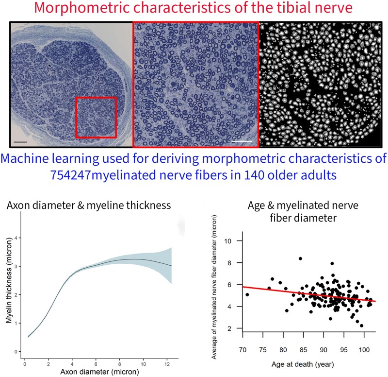
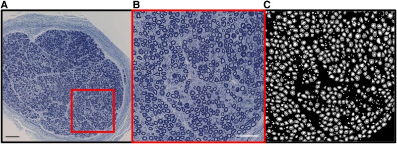
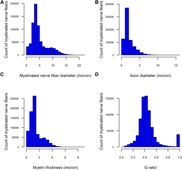
Peripheral nerve comprises a crucial component of the distributed motor/sensory system. However, there is a paucity of data on peripheral nerve morphology derived from large numbers of older adults. This study aimed to quantify the morphometric characteristics of myelinated nerve fibres of the tibial nerve obtained from deceased community-dwelling older adults and examine their association with age. The tibial nerves were obtained from consecutive autopsies of older adults without a history of diabetes who were participants of the Rush Memory and Aging Project, an ongoing longitudinal clinical-autopsy study. A nerve fascicle, obtained from a fixed popliteal segment of the tibial nerve, was separated from the blood vessels and adipose tissue for postmortem examination under an optical microscope. Morphometric characteristics of the myelinated nerve fibres were automatically segmented and quantified using our open-source software AxonDeepSeg. The participants (N = 140) had a mean age of 92.0 years (SD = 5.4) at death, and 72.1% (N = 101) were women. We examined 754 247 myelinated nerve fibres, with an average 5387 (SD = 3436) nerve fibres per participant. The average diameter of myelinated nerve fibres was 4.9 µm (SD = 3.1), axon diameter was 2.0 µm (SD = 1.4), myelin thickness was 1.4 µm (SD = 0.96) and the g-ratio (ratio of axon diameter to myelinated nerve fibre diameter) was 0.45 (SD = 0.17). The relationship between axon diameter and myelin thickness was nonlinear. Myelin was thicker in larger axons up to a diameter of 8 µm, beyond which myelin thickness plateaued. Older age at death was associated with smaller myelinated nerve fibres, smaller axons and thinner myelin. However, age at death was not correlated with myelinated nerve fibre density and was not associated with the average of g-ratio. The association between older age and smaller myelinated nerve fibres was largely attributable to a lower percentage of myelinated nerve fibres >8 µm. We conclude that the smaller tibial myelinated nerve fibres observed in older adults may reflect axonal atrophy rather than degeneration and regeneration of the myelinated nerve fibres. Further research is needed to investigate the pathologies and molecular mechanisms underlying these age-related morphometric changes and their clinical implications in older adults.

 求助内容:
求助内容: 应助结果提醒方式:


