Wanyi Huang, Qiaoqun Ou, Yuanchun Liu, Jiaqi Hu, Lina Wang
{"title":"Effect of myeloid-derived suppressor cells on retinal epithelial cells in retinopathy of prematurity model.","authors":"Wanyi Huang, Qiaoqun Ou, Yuanchun Liu, Jiaqi Hu, Lina Wang","doi":"10.21037/tp-2024-578","DOIUrl":null,"url":null,"abstract":"<p><strong>Background: </strong>Retinopathy of prematurity (ROP) is a proliferative vascular disease with a high incidence rate. Myeloid-derived suppressor cells (MDSCs) are a type of stem cell that possesses remarkable regenerative and reparative capabilities. However, the role of MDSCs as a stem cell therapy in the treatment of ROP remains unclear. Therefore, this study aimed to explore the effects of MDSC therapy on the expression of glial fibrillary acidic protein (GFAP) and vascular endothelial growth factor (VEGF) in a model of ROP, thereby preliminarily assessing the therapeutic potential of MDSCs in ROP.</p><p><strong>Methods: </strong>Bone marrow-derived MDSCs were obtained from an 8-week-old male C57BL/6J mouse, which served as the cell donor. The cells were divided into the model group, which was established by culturing hydrogen peroxide (H<sub>2</sub>O<sub>2</sub>; 300 µM) with adult retinal pigment epithelial cell line-19 (ARPE-19) alone to create the ROP model; the model + medium group having ARPE-19 and medium; and the model + MDSCs group, having ARPE-19 and MDSCs.</p><p><strong>Results: </strong>The expression levels of GFAP and VEGF in the model + MDSCs group were significantly lower compared to the model group. In the model + MDSCs group, GFAP and VEGF expression levels were significantly reduced. Immunofluorescence analysis revealed a significant increase in GFAP and VEGF expression in the model + medium group. In contrast, the expression levels of GFAP and VEGF in the model + MDSCs group were markedly decreased.</p><p><strong>Conclusions: </strong>Co-culturing with MDSCs significantly reduces the expression levels of GFAP and VEGF in the model group, thereby inhibiting pathological neovascularization, inflammatory responses, and immune-mediated damage. This intervention subsequently improves the pathological processes associated with ROP.</p>","PeriodicalId":23294,"journal":{"name":"Translational pediatrics","volume":"14 6","pages":"1147-1155"},"PeriodicalIF":1.7000,"publicationDate":"2025-06-27","publicationTypes":"Journal Article","fieldsOfStudy":null,"isOpenAccess":false,"openAccessPdf":"https://www.ncbi.nlm.nih.gov/pmc/articles/PMC12268656/pdf/","citationCount":"0","resultStr":null,"platform":"Semanticscholar","paperid":null,"PeriodicalName":"Translational pediatrics","FirstCategoryId":"3","ListUrlMain":"https://doi.org/10.21037/tp-2024-578","RegionNum":4,"RegionCategory":"医学","ArticlePicture":[],"TitleCN":null,"AbstractTextCN":null,"PMCID":null,"EPubDate":"2025/6/13 0:00:00","PubModel":"Epub","JCR":"Q2","JCRName":"PEDIATRICS","Score":null,"Total":0}
引用次数: 0
Abstract
Background: Retinopathy of prematurity (ROP) is a proliferative vascular disease with a high incidence rate. Myeloid-derived suppressor cells (MDSCs) are a type of stem cell that possesses remarkable regenerative and reparative capabilities. However, the role of MDSCs as a stem cell therapy in the treatment of ROP remains unclear. Therefore, this study aimed to explore the effects of MDSC therapy on the expression of glial fibrillary acidic protein (GFAP) and vascular endothelial growth factor (VEGF) in a model of ROP, thereby preliminarily assessing the therapeutic potential of MDSCs in ROP.
Methods: Bone marrow-derived MDSCs were obtained from an 8-week-old male C57BL/6J mouse, which served as the cell donor. The cells were divided into the model group, which was established by culturing hydrogen peroxide (H2O2; 300 µM) with adult retinal pigment epithelial cell line-19 (ARPE-19) alone to create the ROP model; the model + medium group having ARPE-19 and medium; and the model + MDSCs group, having ARPE-19 and MDSCs.
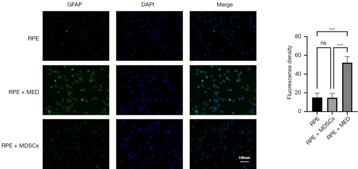
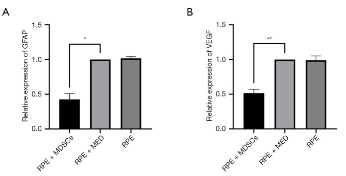
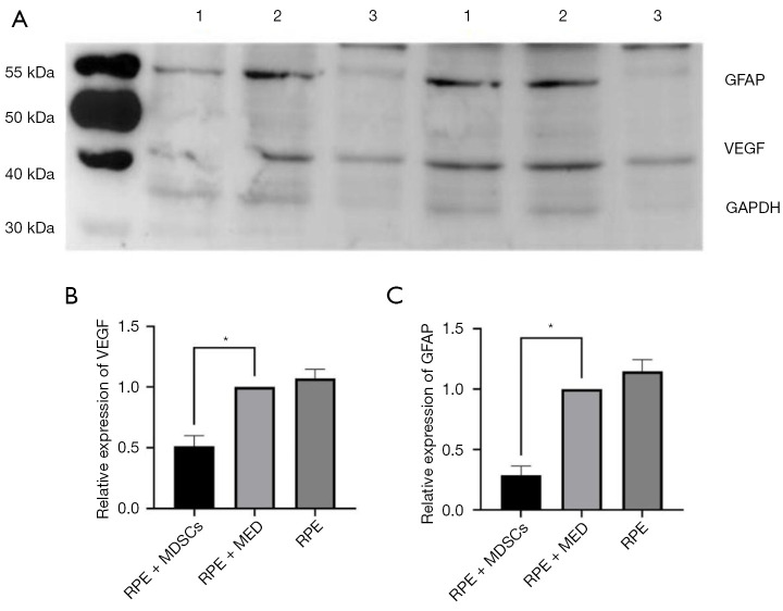
Results: The expression levels of GFAP and VEGF in the model + MDSCs group were significantly lower compared to the model group. In the model + MDSCs group, GFAP and VEGF expression levels were significantly reduced. Immunofluorescence analysis revealed a significant increase in GFAP and VEGF expression in the model + medium group. In contrast, the expression levels of GFAP and VEGF in the model + MDSCs group were markedly decreased.
Conclusions: Co-culturing with MDSCs significantly reduces the expression levels of GFAP and VEGF in the model group, thereby inhibiting pathological neovascularization, inflammatory responses, and immune-mediated damage. This intervention subsequently improves the pathological processes associated with ROP.

 求助内容:
求助内容: 应助结果提醒方式:


