Hong-Bo Xu, Can Ding, Min Zhao, Fa-Jin Lv, Zhi-Gang Chu
{"title":"Exploring the key clinical and CT characteristics of granulomas mimicking peripheral lung cancers: a case-control study.","authors":"Hong-Bo Xu, Can Ding, Min Zhao, Fa-Jin Lv, Zhi-Gang Chu","doi":"10.1186/s13244-025-02043-0","DOIUrl":null,"url":null,"abstract":"<p><strong>Objectives: </strong>Some granulomas exhibit CT manifestations similar to those of peripheral lung cancers (PLCs), often resulting in misdiagnosis. This study aimed to identify the key clinical and CT indicators for differentiating them.</p><p><strong>Materials and methods: </strong>From October 2019 to July 2024, 204 atypical granulomas (no calcification, satellite lesions, and/or halo sign) and 204 size-matched PLCs manifested as solid nodules (SNs) were retrospectively enrolled. Patients' clinical, as well as non-enhanced and contrast-enhanced CT data, were evaluated and compared. The enhancement patterns of lesions included no significant enhancement (▵CT value < 15 HU), rim enhancement, enhancement with well-defined necrosis, heterogeneous enhancement, and homogeneous enhancement. The latter two patterns were further divided into mild (15-29 HU), moderate (30-59 HU), and severe (≥ 60 HU) enhancement.</p><p><strong>Results: </strong>Multivariate analysis revealed that younger age (≤ 63 years) (odds ratio [OR], 5.237; 95% confidence interval [CI], 2.609-10.509; p < 0.001), history of diabetes (OR, 9.097; 95% CI: 3.056-27.077; p < 0.001), irregular shape (OR, 3.603; 95% CI: 1.594-8.142; p = 0.002), lower non-enhanced CT value (≤ 21 HU) (OR, 7.576; 95% CI: 3.720-15.431; p < 0.001), and non-moderate enhancement patterns (OR, 50.065; 95% CI: 20.293-123.517; p < 0.001) were independent predictors of granulomas. The sensitivity, specificity, and area under the curve of this model were 88.7%, 83.8%, and 0.941 (95% CI: 0.919-0.962) (p < 0.001), respectively.</p><p><strong>Conclusions: </strong>In younger (≤ 63 years) patients with diabetes, an irregular SN displaying lower density (≤ 21 HU) in non-enhanced CT and a non-moderate enhancement pattern should first be considered as a granuloma.</p><p><strong>Clinical relevance statement: </strong>Distinguishing atypical granulomas from PLCs can be effectively achieved by evaluating the patient's age, underlying diseases, and the lesion's shape, non-enhanced CT value, and enhancement pattern. This integrated clinical-CT diagnostic approach could provide crucial insights for guiding subsequent clinical management.</p><p><strong>Key points: </strong>Atypical granulomas and PLCs exhibit high morphological similarity. Enhancement patterns of lesions are crucial for differentiating atypical granulomas and PLCs. Atypical granulomas typically display irregular shape, lower non-enhanced CT value, and non-moderate enhancement pattern. Younger age and a history of diabetes are key clinical indicators of granulomas.</p>","PeriodicalId":13639,"journal":{"name":"Insights into Imaging","volume":"16 1","pages":"157"},"PeriodicalIF":4.5000,"publicationDate":"2025-07-19","publicationTypes":"Journal Article","fieldsOfStudy":null,"isOpenAccess":false,"openAccessPdf":"https://www.ncbi.nlm.nih.gov/pmc/articles/PMC12276186/pdf/","citationCount":"0","resultStr":null,"platform":"Semanticscholar","paperid":null,"PeriodicalName":"Insights into Imaging","FirstCategoryId":"3","ListUrlMain":"https://doi.org/10.1186/s13244-025-02043-0","RegionNum":2,"RegionCategory":"医学","ArticlePicture":[],"TitleCN":null,"AbstractTextCN":null,"PMCID":null,"EPubDate":"","PubModel":"","JCR":"Q1","JCRName":"RADIOLOGY, NUCLEAR MEDICINE & MEDICAL IMAGING","Score":null,"Total":0}
引用次数: 0
Abstract
Objectives: Some granulomas exhibit CT manifestations similar to those of peripheral lung cancers (PLCs), often resulting in misdiagnosis. This study aimed to identify the key clinical and CT indicators for differentiating them.
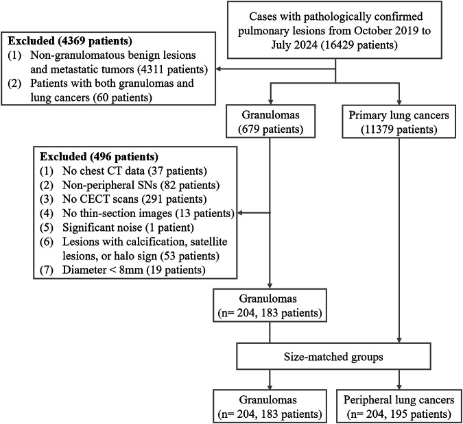
Materials and methods: From October 2019 to July 2024, 204 atypical granulomas (no calcification, satellite lesions, and/or halo sign) and 204 size-matched PLCs manifested as solid nodules (SNs) were retrospectively enrolled. Patients' clinical, as well as non-enhanced and contrast-enhanced CT data, were evaluated and compared. The enhancement patterns of lesions included no significant enhancement (▵CT value < 15 HU), rim enhancement, enhancement with well-defined necrosis, heterogeneous enhancement, and homogeneous enhancement. The latter two patterns were further divided into mild (15-29 HU), moderate (30-59 HU), and severe (≥ 60 HU) enhancement.
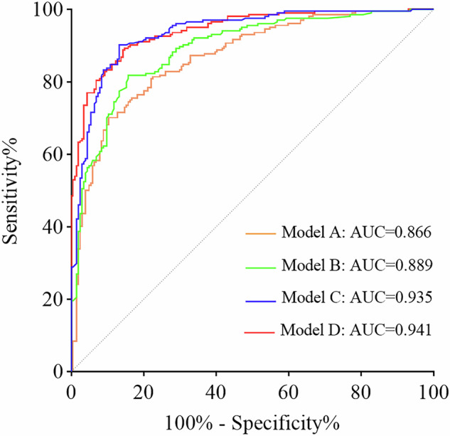
Results: Multivariate analysis revealed that younger age (≤ 63 years) (odds ratio [OR], 5.237; 95% confidence interval [CI], 2.609-10.509; p < 0.001), history of diabetes (OR, 9.097; 95% CI: 3.056-27.077; p < 0.001), irregular shape (OR, 3.603; 95% CI: 1.594-8.142; p = 0.002), lower non-enhanced CT value (≤ 21 HU) (OR, 7.576; 95% CI: 3.720-15.431; p < 0.001), and non-moderate enhancement patterns (OR, 50.065; 95% CI: 20.293-123.517; p < 0.001) were independent predictors of granulomas. The sensitivity, specificity, and area under the curve of this model were 88.7%, 83.8%, and 0.941 (95% CI: 0.919-0.962) (p < 0.001), respectively.
Conclusions: In younger (≤ 63 years) patients with diabetes, an irregular SN displaying lower density (≤ 21 HU) in non-enhanced CT and a non-moderate enhancement pattern should first be considered as a granuloma.
Clinical relevance statement: Distinguishing atypical granulomas from PLCs can be effectively achieved by evaluating the patient's age, underlying diseases, and the lesion's shape, non-enhanced CT value, and enhancement pattern. This integrated clinical-CT diagnostic approach could provide crucial insights for guiding subsequent clinical management.
Key points: Atypical granulomas and PLCs exhibit high morphological similarity. Enhancement patterns of lesions are crucial for differentiating atypical granulomas and PLCs. Atypical granulomas typically display irregular shape, lower non-enhanced CT value, and non-moderate enhancement pattern. Younger age and a history of diabetes are key clinical indicators of granulomas.
期刊介绍:
Insights into Imaging (I³) is a peer-reviewed open access journal published under the brand SpringerOpen. All content published in the journal is freely available online to anyone, anywhere!
I³ continuously updates scientific knowledge and progress in best-practice standards in radiology through the publication of original articles and state-of-the-art reviews and opinions, along with recommendations and statements from the leading radiological societies in Europe.
Founded by the European Society of Radiology (ESR), I³ creates a platform for educational material, guidelines and recommendations, and a forum for topics of controversy.
A balanced combination of review articles, original papers, short communications from European radiological congresses and information on society matters makes I³ an indispensable source for current information in this field.
I³ is owned by the ESR, however authors retain copyright to their article according to the Creative Commons Attribution License (see Copyright and License Agreement). All articles can be read, redistributed and reused for free, as long as the author of the original work is cited properly.
The open access fees (article-processing charges) for this journal are kindly sponsored by ESR for all Members.
The journal went open access in 2012, which means that all articles published since then are freely available online.

 求助内容:
求助内容: 应助结果提醒方式:


