Markus Kist, Paul Strenge, Tobias Keck, Andreas Weber, Peter Bronsert, Thaer S A Abdalla, Ulrich Friedrich Wellner, Michael Thomaschewski
{"title":"Intraoperative differentiation of pancreatic neoplastic lesions using optical coherence tomography (OCT).","authors":"Markus Kist, Paul Strenge, Tobias Keck, Andreas Weber, Peter Bronsert, Thaer S A Abdalla, Ulrich Friedrich Wellner, Michael Thomaschewski","doi":"10.1007/s00423-025-03810-9","DOIUrl":null,"url":null,"abstract":"<p><strong>Purpose: </strong>The diagnostic methods for accurately differentiating the dignity of pancreatic neoplasms are limited. Worrisome features on MRI and endosonography guide the way to resection or conservative treatment with a relevant rate of failure. Intraoperative minimal invasive optical coherence tomography could be a solution for this challenge. The aim of this study is to investigate whether optical coherence tomography is suitable for differentiating of pancreatic neoplastic lesions.</p><p><strong>Methods: </strong>In this exploratory study, four patient's specimens of pancreatic resections (white adipose tissue, intraductal papillary mucinous neoplasm (IPMN), pancreatic ductal adenocarcinoma (PDAC) based on IPMN and neuroendocrine pancreatic carcinoma) were prospectively examined ex vivo immediately after resection in the operating room using an optical coherence tomography system (Callisto 930nm, Thorlabs GmbH). In detail, the study investigated whether and in what way endocrine tumors, adenocarcinomas, premalignant and benign cysts differ morphologically in optical coherence tomography imaging compared to healthy pancreatic tissue. The final histopathological findings of the pancreatic specimens served as a reference and were correlated.</p><p><strong>Results: </strong>The samples examined ranged from typical fatty tissue, intraductal papillary mucinous neoplasm (IPMN), a moderate differentiated (G2) pancreatic ductal adenocarcinoma (PDAC) based on an intraductal papillary mucinous neoplasm (IPMN) and a neuroendocrine pancreatic carcinoma. Optical coherence tomography was feasible to replicate key histological characteristics and tissue architecture in correlation to conventional Hematoxylin-eosin histology.</p><p><strong>Conclusion: </strong>Optical coherence tomography imaging has the potential to differentiate between benign, pre-malignant and malignant pancreatic pathologies by morphology and should be examined in larger collectives.</p>","PeriodicalId":17983,"journal":{"name":"Langenbeck's Archives of Surgery","volume":"410 1","pages":"227"},"PeriodicalIF":1.8000,"publicationDate":"2025-07-18","publicationTypes":"Journal Article","fieldsOfStudy":null,"isOpenAccess":false,"openAccessPdf":"https://www.ncbi.nlm.nih.gov/pmc/articles/PMC12274226/pdf/","citationCount":"0","resultStr":null,"platform":"Semanticscholar","paperid":null,"PeriodicalName":"Langenbeck's Archives of Surgery","FirstCategoryId":"3","ListUrlMain":"https://doi.org/10.1007/s00423-025-03810-9","RegionNum":3,"RegionCategory":"医学","ArticlePicture":[],"TitleCN":null,"AbstractTextCN":null,"PMCID":null,"EPubDate":"","PubModel":"","JCR":"Q2","JCRName":"SURGERY","Score":null,"Total":0}
引用次数: 0
Abstract
Purpose: The diagnostic methods for accurately differentiating the dignity of pancreatic neoplasms are limited. Worrisome features on MRI and endosonography guide the way to resection or conservative treatment with a relevant rate of failure. Intraoperative minimal invasive optical coherence tomography could be a solution for this challenge. The aim of this study is to investigate whether optical coherence tomography is suitable for differentiating of pancreatic neoplastic lesions.
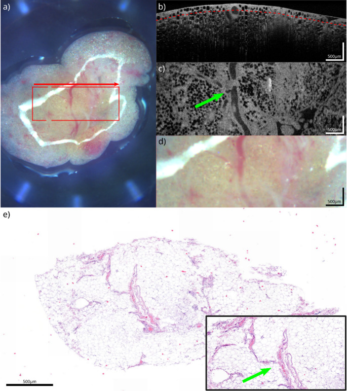
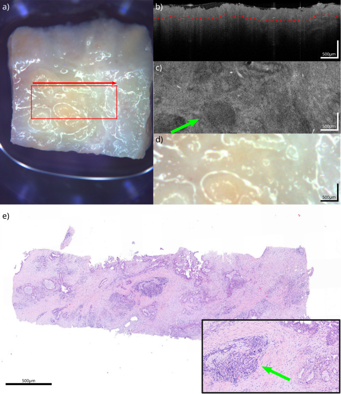
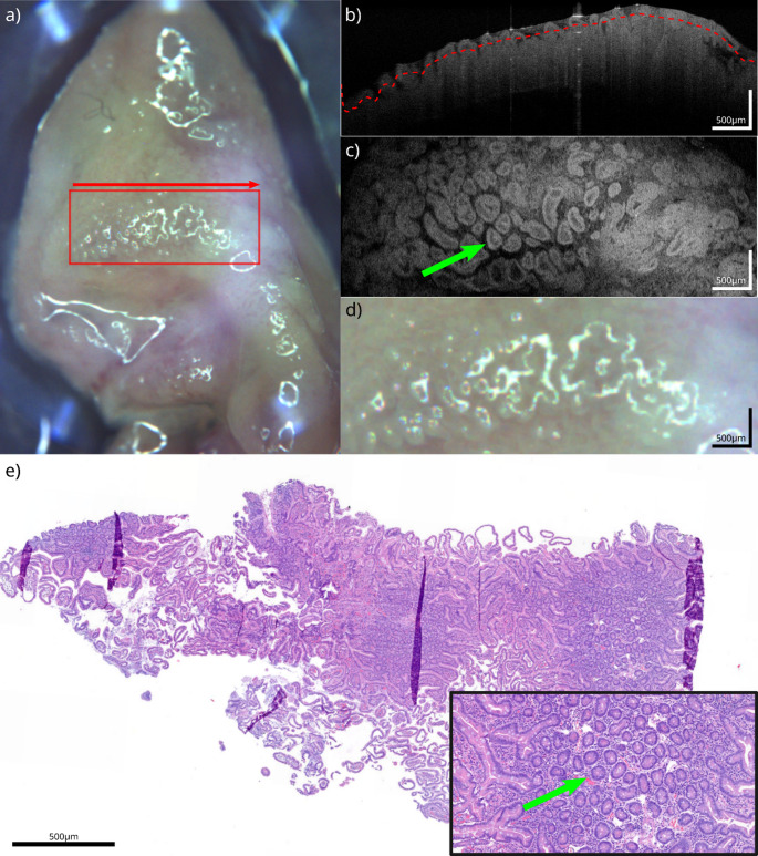
Methods: In this exploratory study, four patient's specimens of pancreatic resections (white adipose tissue, intraductal papillary mucinous neoplasm (IPMN), pancreatic ductal adenocarcinoma (PDAC) based on IPMN and neuroendocrine pancreatic carcinoma) were prospectively examined ex vivo immediately after resection in the operating room using an optical coherence tomography system (Callisto 930nm, Thorlabs GmbH). In detail, the study investigated whether and in what way endocrine tumors, adenocarcinomas, premalignant and benign cysts differ morphologically in optical coherence tomography imaging compared to healthy pancreatic tissue. The final histopathological findings of the pancreatic specimens served as a reference and were correlated.
Results: The samples examined ranged from typical fatty tissue, intraductal papillary mucinous neoplasm (IPMN), a moderate differentiated (G2) pancreatic ductal adenocarcinoma (PDAC) based on an intraductal papillary mucinous neoplasm (IPMN) and a neuroendocrine pancreatic carcinoma. Optical coherence tomography was feasible to replicate key histological characteristics and tissue architecture in correlation to conventional Hematoxylin-eosin histology.
Conclusion: Optical coherence tomography imaging has the potential to differentiate between benign, pre-malignant and malignant pancreatic pathologies by morphology and should be examined in larger collectives.
期刊介绍:
Langenbeck''s Archives of Surgery aims to publish the best results in the field of clinical surgery and basic surgical research. The main focus is on providing the highest level of clinical research and clinically relevant basic research. The journal, published exclusively in English, will provide an international discussion forum for the controlled results of clinical surgery. The majority of published contributions will be original articles reporting on clinical data from general and visceral surgery, while endocrine surgery will also be covered. Papers on basic surgical principles from the fields of traumatology, vascular and thoracic surgery are also welcome. Evidence-based medicine is an important criterion for the acceptance of papers.
 求助内容:
求助内容: 应助结果提醒方式:


