{"title":"Deep learning reconstruction enhances image quality in contrast-enhanced CT venography for deep vein thrombosis.","authors":"Yusuke Asari, Koichiro Yasaka, Joji Kurashima, Akira Katayama, Mariko Kurokawa, Osamu Abe","doi":"10.1007/s10140-025-02366-x","DOIUrl":null,"url":null,"abstract":"<p><strong>Purpose: </strong>This study aimed to evaluate and compare the diagnostic performance and image quality of deep learning reconstruction (DLR) with hybrid iterative reconstruction (Hybrid IR) and filtered back projection (FBP) in contrast-enhanced CT venography for deep vein thrombosis (DVT).</p><p><strong>Methods: </strong>A retrospective analysis was conducted on 51 patients who underwent lower limb CT venography, including 20 with DVT lesions and 31 without DVT lesions. CT images were reconstructed using DLR, Hybrid IR, and FBP. Quantitative image quality metrics, such as contrast-to-noise ratio (CNR) and image noise, were measured. Three radiologists independently assessed DVT lesion detection, depiction of DVT lesions and normal structures, subjective image noise, artifacts, and overall image quality using scoring systems. Diagnostic performance was evaluated using sensitivity and area under the receiver operating characteristic curve (AUC). The paired t-test and Wilcoxon signed-rank test compared the results for continuous variables and ordinal scales, respectively, between DLR and Hybrid IR as well as between DLR and FBP.</p><p><strong>Results: </strong>DLR significantly improved CNR and reduced image noise compared to Hybrid IR and FBP (p < 0.001). AUC and sensitivity for DVT detection were not statistically different across reconstruction methods. Two readers reported improved lesion visualization with DLR. DLR was also rated superior in image quality, normal structure depiction, and noise suppression by all readers (p < 0.001).</p><p><strong>Conclusions: </strong>DLR enhances image quality and anatomical clarity in CT venography. These findings support the utility of DLR in improving diagnostic confidence and image interpretability in DVT assessment.</p>","PeriodicalId":11623,"journal":{"name":"Emergency Radiology","volume":" ","pages":"699-708"},"PeriodicalIF":1.3000,"publicationDate":"2025-10-01","publicationTypes":"Journal Article","fieldsOfStudy":null,"isOpenAccess":false,"openAccessPdf":"https://www.ncbi.nlm.nih.gov/pmc/articles/PMC12496264/pdf/","citationCount":"0","resultStr":null,"platform":"Semanticscholar","paperid":null,"PeriodicalName":"Emergency Radiology","FirstCategoryId":"1085","ListUrlMain":"https://doi.org/10.1007/s10140-025-02366-x","RegionNum":0,"RegionCategory":null,"ArticlePicture":[],"TitleCN":null,"AbstractTextCN":null,"PMCID":null,"EPubDate":"2025/7/18 0:00:00","PubModel":"Epub","JCR":"Q3","JCRName":"RADIOLOGY, NUCLEAR MEDICINE & MEDICAL IMAGING","Score":null,"Total":0}
引用次数: 0
Abstract
Purpose: This study aimed to evaluate and compare the diagnostic performance and image quality of deep learning reconstruction (DLR) with hybrid iterative reconstruction (Hybrid IR) and filtered back projection (FBP) in contrast-enhanced CT venography for deep vein thrombosis (DVT).
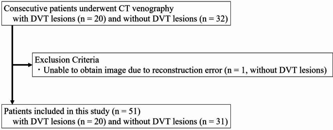
Methods: A retrospective analysis was conducted on 51 patients who underwent lower limb CT venography, including 20 with DVT lesions and 31 without DVT lesions. CT images were reconstructed using DLR, Hybrid IR, and FBP. Quantitative image quality metrics, such as contrast-to-noise ratio (CNR) and image noise, were measured. Three radiologists independently assessed DVT lesion detection, depiction of DVT lesions and normal structures, subjective image noise, artifacts, and overall image quality using scoring systems. Diagnostic performance was evaluated using sensitivity and area under the receiver operating characteristic curve (AUC). The paired t-test and Wilcoxon signed-rank test compared the results for continuous variables and ordinal scales, respectively, between DLR and Hybrid IR as well as between DLR and FBP.
Results: DLR significantly improved CNR and reduced image noise compared to Hybrid IR and FBP (p < 0.001). AUC and sensitivity for DVT detection were not statistically different across reconstruction methods. Two readers reported improved lesion visualization with DLR. DLR was also rated superior in image quality, normal structure depiction, and noise suppression by all readers (p < 0.001).
Conclusions: DLR enhances image quality and anatomical clarity in CT venography. These findings support the utility of DLR in improving diagnostic confidence and image interpretability in DVT assessment.
期刊介绍:
To advance and improve the radiologic aspects of emergency careTo establish Emergency Radiology as an area of special interest in the field of diagnostic imagingTo improve methods of education in Emergency RadiologyTo provide, through formal meetings, a mechanism for presentation of scientific papers on various aspects of Emergency Radiology and continuing educationTo promote research in Emergency Radiology by clinical and basic science investigators, including residents and other traineesTo act as the resource body on Emergency Radiology for those interested in emergency patient care Members of the American Society of Emergency Radiology (ASER) receive the Emergency Radiology journal as a benefit of membership!


 求助内容:
求助内容: 应助结果提醒方式:


