Thyroid tuberculosis misdiagnosed as papillary thyroid carcinoma under ultrasound-guided fine-needle aspiration cytology: a case report and literature review.
Lu Gan, Li Sun, Junzhi Zhao, Qiang Feng, Jiahua Li, Qinyun Wan, Qiannan Meng, Jianxue Liu
{"title":"Thyroid tuberculosis misdiagnosed as papillary thyroid carcinoma under ultrasound-guided fine-needle aspiration cytology: a case report and literature review.","authors":"Lu Gan, Li Sun, Junzhi Zhao, Qiang Feng, Jiahua Li, Qinyun Wan, Qiannan Meng, Jianxue Liu","doi":"10.21037/gs-2025-19","DOIUrl":null,"url":null,"abstract":"<p><strong>Background: </strong>Thyroid tuberculosis is very rare, making diagnosis challenging without fine-needle aspiration cytology (FNAC) because of its atypical features. We report a case of thyroid tuberculosis that was misdiagnosed as papillary thyroid carcinoma (PTC).</p><p><strong>Case description: </strong>A 68-year-old woman visited Baoji Central Hospital following ultrasound (US) performed at another hospital indicated a suspicious malignant nodule in her thyroid. Physical examination and thyroid US revealed a hard mass and irregular hypoechoic area in the right lobe, suggesting a possible malignant lesion. Ultrasound-guided FNAC (US-FNAC) was suspicious for PTC. The patient subsequently underwent right thyroid lobectomy and isthmusectomy, and postoperative histopathology revealed an epithelioid granulomatous lesion without tumor tissue. Meanwhile, postoperative serum testing revealed elevated TB-γ interferon concentration, and a specific T lymphocyte testing was positive, indicating a tuberculosis infection. Primary thyroid tuberculosis was ultimately confirmed. Postoperatively, the patient recovered well after surgery and received anti-tuberculosis therapy in a tuberculosis hospital for 1 year.</p><p><strong>Conclusions: </strong>This case reminds us that although thyroid tuberculosis is very rare, especially when imaging features and FNAC results resemble those of PTC, the diagnosis of primary thyroid tuberculosis should still be considered, and serum TB-related indicator testing can aid in diagnosis.</p>","PeriodicalId":12760,"journal":{"name":"Gland surgery","volume":"14 6","pages":"1154-1160"},"PeriodicalIF":1.6000,"publicationDate":"2025-06-30","publicationTypes":"Journal Article","fieldsOfStudy":null,"isOpenAccess":false,"openAccessPdf":"https://www.ncbi.nlm.nih.gov/pmc/articles/PMC12261367/pdf/","citationCount":"0","resultStr":null,"platform":"Semanticscholar","paperid":null,"PeriodicalName":"Gland surgery","FirstCategoryId":"3","ListUrlMain":"https://doi.org/10.21037/gs-2025-19","RegionNum":3,"RegionCategory":"医学","ArticlePicture":[],"TitleCN":null,"AbstractTextCN":null,"PMCID":null,"EPubDate":"2025/6/26 0:00:00","PubModel":"Epub","JCR":"Q3","JCRName":"SURGERY","Score":null,"Total":0}
引用次数: 0
Abstract
Background: Thyroid tuberculosis is very rare, making diagnosis challenging without fine-needle aspiration cytology (FNAC) because of its atypical features. We report a case of thyroid tuberculosis that was misdiagnosed as papillary thyroid carcinoma (PTC).
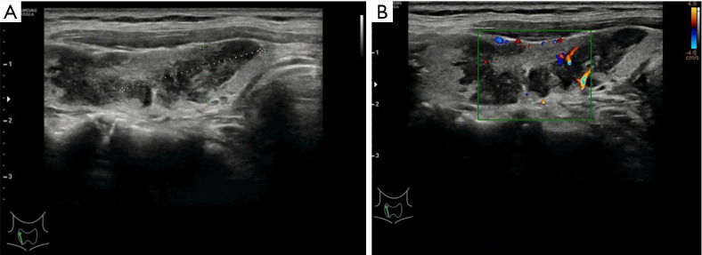
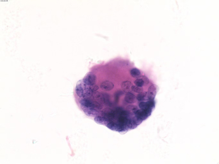
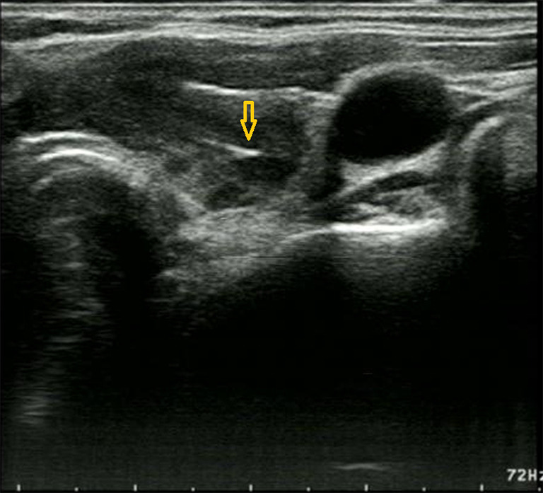
Case description: A 68-year-old woman visited Baoji Central Hospital following ultrasound (US) performed at another hospital indicated a suspicious malignant nodule in her thyroid. Physical examination and thyroid US revealed a hard mass and irregular hypoechoic area in the right lobe, suggesting a possible malignant lesion. Ultrasound-guided FNAC (US-FNAC) was suspicious for PTC. The patient subsequently underwent right thyroid lobectomy and isthmusectomy, and postoperative histopathology revealed an epithelioid granulomatous lesion without tumor tissue. Meanwhile, postoperative serum testing revealed elevated TB-γ interferon concentration, and a specific T lymphocyte testing was positive, indicating a tuberculosis infection. Primary thyroid tuberculosis was ultimately confirmed. Postoperatively, the patient recovered well after surgery and received anti-tuberculosis therapy in a tuberculosis hospital for 1 year.
Conclusions: This case reminds us that although thyroid tuberculosis is very rare, especially when imaging features and FNAC results resemble those of PTC, the diagnosis of primary thyroid tuberculosis should still be considered, and serum TB-related indicator testing can aid in diagnosis.
期刊介绍:
Gland Surgery (Gland Surg; GS, Print ISSN 2227-684X; Online ISSN 2227-8575) being indexed by PubMed/PubMed Central, is an open access, peer-review journal launched at May of 2012, published bio-monthly since February 2015.
 求助内容:
求助内容: 应助结果提醒方式:


