Bisharah Rizvi, Jorge A Munoz Pineda, Keriann Van Nostrand, Russell Miller, George Cheng, Niral M Patel
{"title":"Complications of linear endobronchial ultrasound guided biopsies: narrative review.","authors":"Bisharah Rizvi, Jorge A Munoz Pineda, Keriann Van Nostrand, Russell Miller, George Cheng, Niral M Patel","doi":"10.21037/med-24-33","DOIUrl":null,"url":null,"abstract":"<p><strong>Background and objective: </strong>Linear endobronchial ultrasound (EBUS) has become a key tool for diagnosing pulmonary diseases, offering high diagnostic yield for both malignant and non-malignant conditions. With its increased use, more complications are being reported. The objective of this narrative review is to discuss the complications associated with linear EBUS.</p><p><strong>Methods: </strong>A literature search using PubMed and Google Scholar from 2009 to 2024 was done. We included case reports, prospective, and retrospective studies reporting linear EBUS complications.</p><p><strong>Key content and findings: </strong>Overall complications from EBUS range from 0.04% to 17%. Most common are infectious complications which are 0.04-4%. These include mediastinitis, pneumonia, pericarditis, bacteremia, tumor bed infection, lung abscess, empyema, and septic shock. Other complications include pneumothorax, pneumomediastinum, pneumopericardium, pneumoperitoneum, and subcutaneous emphysema. Complications due to anesthesia or equipment malfunction can occur as well. Hemorrhagic complications have been reported as well. Mortality is low 0.01-0.04%, and four cases have been reported that led to death from complications.</p><p><strong>Conclusions: </strong>With increased use of EBUS as a diagnostic tool, number of complications will increase. Clinicians performing the procedures should be aware of types of possible complications that can occur and follow the patients closely after the procedure. Rapid diagnosis and treatment should be done to avoid fatal outcomes.</p>","PeriodicalId":74139,"journal":{"name":"Mediastinum (Hong Kong, China)","volume":"9 ","pages":"12"},"PeriodicalIF":0.0000,"publicationDate":"2025-05-21","publicationTypes":"Journal Article","fieldsOfStudy":null,"isOpenAccess":false,"openAccessPdf":"https://www.ncbi.nlm.nih.gov/pmc/articles/PMC12260959/pdf/","citationCount":"0","resultStr":null,"platform":"Semanticscholar","paperid":null,"PeriodicalName":"Mediastinum (Hong Kong, China)","FirstCategoryId":"1085","ListUrlMain":"https://doi.org/10.21037/med-24-33","RegionNum":0,"RegionCategory":null,"ArticlePicture":[],"TitleCN":null,"AbstractTextCN":null,"PMCID":null,"EPubDate":"2025/1/1 0:00:00","PubModel":"eCollection","JCR":"","JCRName":"","Score":null,"Total":0}
引用次数: 0
Abstract
Background and objective: Linear endobronchial ultrasound (EBUS) has become a key tool for diagnosing pulmonary diseases, offering high diagnostic yield for both malignant and non-malignant conditions. With its increased use, more complications are being reported. The objective of this narrative review is to discuss the complications associated with linear EBUS.
Methods: A literature search using PubMed and Google Scholar from 2009 to 2024 was done. We included case reports, prospective, and retrospective studies reporting linear EBUS complications.
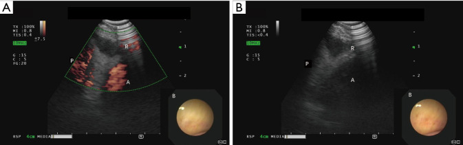
Key content and findings: Overall complications from EBUS range from 0.04% to 17%. Most common are infectious complications which are 0.04-4%. These include mediastinitis, pneumonia, pericarditis, bacteremia, tumor bed infection, lung abscess, empyema, and septic shock. Other complications include pneumothorax, pneumomediastinum, pneumopericardium, pneumoperitoneum, and subcutaneous emphysema. Complications due to anesthesia or equipment malfunction can occur as well. Hemorrhagic complications have been reported as well. Mortality is low 0.01-0.04%, and four cases have been reported that led to death from complications.
Conclusions: With increased use of EBUS as a diagnostic tool, number of complications will increase. Clinicians performing the procedures should be aware of types of possible complications that can occur and follow the patients closely after the procedure. Rapid diagnosis and treatment should be done to avoid fatal outcomes.

 求助内容:
求助内容: 应助结果提醒方式:


