Denise S Hoogenkamp, Berlinda J de Wit-van der Veen, Jeske Hendriksen, Karlijn van der Schilden, Josefine A M Nanne, José S A Belderbos, Maddalena M Rossi, Martien P J Mooijer, Uta Funke, N Harry Hendrikse, Wouter V Vogel, Else A Aalbersberg
{"title":"[<sup>195m</sup>Pt]Cisplatin for lung cancer imaging: a pilot study.","authors":"Denise S Hoogenkamp, Berlinda J de Wit-van der Veen, Jeske Hendriksen, Karlijn van der Schilden, Josefine A M Nanne, José S A Belderbos, Maddalena M Rossi, Martien P J Mooijer, Uta Funke, N Harry Hendrikse, Wouter V Vogel, Else A Aalbersberg","doi":"10.1186/s13550-025-01281-z","DOIUrl":null,"url":null,"abstract":"<p><strong>Background: </strong>Radiolabeled [<sup>195m</sup>Pt]cisplatin has been developed as a tool to determine the biodistribution of cisplatin in vivo through SPECT imaging and possibly aid in patient-selection. This feasibility study aimed to evaluate the (radiation) safety, biodistribution, and image quality of [<sup>195m</sup>Pt]cisplatin SPECT/CT in patients with non-small cell lung cancer (NSCLC). [<sup>195m</sup>Pt]Cisplatin was produced under GMP standards. Six patients received 100 MBq [<sup>195m</sup>Pt]cisplatin in their second or third week of chemoradiation therapy. Planar and SPECT/CT imaging were acquired at 1.5, 48, 120 and 168 h post-administration. Segmentation on SPECT for biodistribution and dosimetry was achieved using TotalSegmentator in 3DSlicer, and organ specific time-activity curves were generated through a mono-exponential curve fit. Effective and absorbed doses were obtained with S-values from IDAC-Dose 2.1. Toxicity was assessed using CTCAE criteria.</p><p><strong>Results: </strong>Six NSCLC patients received 100.9 ± 3.3 MBq [<sup>195m</sup>Pt]cisplatin at a radioactivity concentration of 11.1 ± 4.9 MBq/mL. No adverse events above grade 2 were observed. [<sup>195m</sup>Pt]Cisplatin had a long retention time with an effective half-life of 74.8 h. Uptake was highest in the liver and kidneys, which also resulted in the highest absorbed dose at 48.6 ± 7.9 mGy and 38.4 ± 7.3 mGy, respectively. Tumor uptake was similar to blood, with a ratio of 1.0 ± 0.05.</p><p><strong>Conclusion: </strong>[<sup>195m</sup>Pt]Cisplatin is safe to use for imaging in patients with NSCLC when injecting 100 MBq, reaching a mean effective dose of 14.6 ± 1.5 mSv. The image quality of [<sup>195m</sup>Pt]cisplatin was suitable for SPECT imaging and quantification. Tumor uptake was similar to blood.</p><p><strong>Trial registration: </strong>CCMO, NL74272.031.21. Registered 28 January 2022, https://www.onderzoekmetmensen.nl/nl/trial/55147 .</p>","PeriodicalId":11611,"journal":{"name":"EJNMMI Research","volume":"15 1","pages":"87"},"PeriodicalIF":3.1000,"publicationDate":"2025-07-16","publicationTypes":"Journal Article","fieldsOfStudy":null,"isOpenAccess":false,"openAccessPdf":"https://www.ncbi.nlm.nih.gov/pmc/articles/PMC12267755/pdf/","citationCount":"0","resultStr":null,"platform":"Semanticscholar","paperid":null,"PeriodicalName":"EJNMMI Research","FirstCategoryId":"3","ListUrlMain":"https://doi.org/10.1186/s13550-025-01281-z","RegionNum":3,"RegionCategory":"医学","ArticlePicture":[],"TitleCN":null,"AbstractTextCN":null,"PMCID":null,"EPubDate":"","PubModel":"","JCR":"Q1","JCRName":"RADIOLOGY, NUCLEAR MEDICINE & MEDICAL IMAGING","Score":null,"Total":0}
引用次数: 0
Abstract
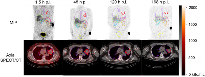
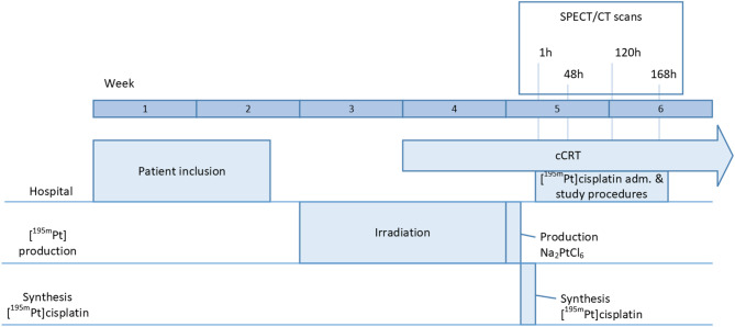
Background: Radiolabeled [195mPt]cisplatin has been developed as a tool to determine the biodistribution of cisplatin in vivo through SPECT imaging and possibly aid in patient-selection. This feasibility study aimed to evaluate the (radiation) safety, biodistribution, and image quality of [195mPt]cisplatin SPECT/CT in patients with non-small cell lung cancer (NSCLC). [195mPt]Cisplatin was produced under GMP standards. Six patients received 100 MBq [195mPt]cisplatin in their second or third week of chemoradiation therapy. Planar and SPECT/CT imaging were acquired at 1.5, 48, 120 and 168 h post-administration. Segmentation on SPECT for biodistribution and dosimetry was achieved using TotalSegmentator in 3DSlicer, and organ specific time-activity curves were generated through a mono-exponential curve fit. Effective and absorbed doses were obtained with S-values from IDAC-Dose 2.1. Toxicity was assessed using CTCAE criteria.
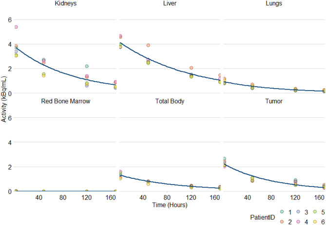
Results: Six NSCLC patients received 100.9 ± 3.3 MBq [195mPt]cisplatin at a radioactivity concentration of 11.1 ± 4.9 MBq/mL. No adverse events above grade 2 were observed. [195mPt]Cisplatin had a long retention time with an effective half-life of 74.8 h. Uptake was highest in the liver and kidneys, which also resulted in the highest absorbed dose at 48.6 ± 7.9 mGy and 38.4 ± 7.3 mGy, respectively. Tumor uptake was similar to blood, with a ratio of 1.0 ± 0.05.
Conclusion: [195mPt]Cisplatin is safe to use for imaging in patients with NSCLC when injecting 100 MBq, reaching a mean effective dose of 14.6 ± 1.5 mSv. The image quality of [195mPt]cisplatin was suitable for SPECT imaging and quantification. Tumor uptake was similar to blood.
Trial registration: CCMO, NL74272.031.21. Registered 28 January 2022, https://www.onderzoekmetmensen.nl/nl/trial/55147 .
EJNMMI ResearchRADIOLOGY, NUCLEAR MEDICINE & MEDICAL IMAGING&nb-
CiteScore
5.90
自引率
3.10%
发文量
72
审稿时长
13 weeks
期刊介绍:
EJNMMI Research publishes new basic, translational and clinical research in the field of nuclear medicine and molecular imaging. Regular features include original research articles, rapid communication of preliminary data on innovative research, interesting case reports, editorials, and letters to the editor. Educational articles on basic sciences, fundamental aspects and controversy related to pre-clinical and clinical research or ethical aspects of research are also welcome. Timely reviews provide updates on current applications, issues in imaging research and translational aspects of nuclear medicine and molecular imaging technologies.
The main emphasis is placed on the development of targeted imaging with radiopharmaceuticals within the broader context of molecular probes to enhance understanding and characterisation of the complex biological processes underlying disease and to develop, test and guide new treatment modalities, including radionuclide therapy.

 求助内容:
求助内容: 应助结果提醒方式:


