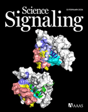
Autophosphorylation of oncoprotein TEL-ABL in myeloid and lymphoid cells confers resistance to the allosteric ABL inhibitor asciminib
IF 6.6
1区 生物学
Q1 BIOCHEMISTRY & MOLECULAR BIOLOGY
Science Signaling
Pub Date : 2025-07-15
引用次数: 0
Abstract
Chromosomal translocations that fuse ABL1 to BCR or TEL cause human leukemias. In BCR-ABL and TEL-ABL fusion proteins, oligomerization and loss of an autoinhibitory myristoylation site in the SH3 domain of ABL lead to increased ABL tyrosine kinase activity. We assessed the ability of asciminib, an allosteric inhibitor of BCR-ABL that binds to the myristoyl-binding site in the ABL kinase domain, to inhibit these fusion proteins. Although the ABL components of the two fusion proteins have identical sequences, asciminib was much less effective against TEL-ABL than it was against BCR-ABL in cell-growth assays. In contrast, ATP-competitive tyrosine kinase inhibitors, such as imatinib and ponatinib, were equally effective against both fusion proteins. A helix in the ABL kinase domain that closes over bound asciminib was required for the sensitivity of BCR-ABL to the drug but had no effect on that of TEL-ABL, suggesting that the native autoinhibitory mechanism that asciminib engages in BCR-ABL is disrupted in TEL-ABL. Single-molecule microscopy demonstrated that BCR-ABL was mainly dimeric in cells, whereas TEL-ABL formed higher-order oligomers, which promoted trans-autophosphorylation, including of a regulatory phosphorylation site (Tyr89) in the SH3 domain of ABL. Nonphosphorylated TEL-ABL was intrinsically susceptible to inhibition by asciminib, but phosphorylation at Tyr89 disassembled the autoinhibited conformation of ABL, thereby preventing asciminib from binding. Our results demonstrate that phosphorylation determines whether an ABL fusion protein is sensitive to allosteric inhibition.
髓细胞和淋巴细胞中癌蛋白TEL-ABL的自磷酸化赋予对变构ABL抑制剂阿西米尼的抗性
将ABL1与BCR或TEL融合的染色体易位导致人类白血病。在BCR-ABL和TEL-ABL融合蛋白中,ABL SH3结构域的自抑制肉豆肉酰化位点的寡聚化和缺失导致ABL酪氨酸激酶活性增加。我们评估了阿西米尼(asciminib)抑制这些融合蛋白的能力,阿西米尼是一种BCR-ABL的变构抑制剂,与ABL激酶结构域的肉豆基结合位点结合。虽然两种融合蛋白的ABL成分具有相同的序列,但在细胞生长试验中,阿西米尼对TEL-ABL的作用远低于对BCR-ABL的作用。相比之下,atp竞争酪氨酸激酶抑制剂,如伊马替尼和波纳替尼,对两种融合蛋白同样有效。ABL激酶结构域的螺旋结构关闭过结合的阿西米尼是BCR-ABL对该药的敏感性所必需的,但对TEL-ABL的敏感性没有影响,这表明阿西米尼参与BCR-ABL的天然自身抑制机制在TEL-ABL中被破坏。单分子显微镜显示,BCR-ABL在细胞中主要是二聚体,而TEL-ABL形成高阶低聚物,促进反式自磷酸化,包括ABL SH3结构域的调控磷酸化位点(Tyr89)。非磷酸化的TEL-ABL本质上容易受到阿西米尼的抑制,但Tyr89位点的磷酸化破坏了ABL的自抑制构象,从而阻止了阿西米尼的结合。我们的研究结果表明,磷酸化决定了ABL融合蛋白是否对变构抑制敏感。
本文章由计算机程序翻译,如有差异,请以英文原文为准。
求助全文
约1分钟内获得全文
求助全文
来源期刊

Science Signaling
BIOCHEMISTRY & MOLECULAR BIOLOGY-CELL BIOLOGY
CiteScore
9.50
自引率
0.00%
发文量
148
审稿时长
3-8 weeks
期刊介绍:
"Science Signaling" is a reputable, peer-reviewed journal dedicated to the exploration of cell communication mechanisms, offering a comprehensive view of the intricate processes that govern cellular regulation. This journal, published weekly online by the American Association for the Advancement of Science (AAAS), is a go-to resource for the latest research in cell signaling and its various facets.
The journal's scope encompasses a broad range of topics, including the study of signaling networks, synthetic biology, systems biology, and the application of these findings in drug discovery. It also delves into the computational and modeling aspects of regulatory pathways, providing insights into how cells communicate and respond to their environment.
In addition to publishing full-length articles that report on groundbreaking research, "Science Signaling" also features reviews that synthesize current knowledge in the field, focus articles that highlight specific areas of interest, and editor-written highlights that draw attention to particularly significant studies. This mix of content ensures that the journal serves as a valuable resource for both researchers and professionals looking to stay abreast of the latest advancements in cell communication science.
 求助内容:
求助内容: 应助结果提醒方式:


