Luka Manojlović, Matija Mamić, Karolina Krstanac, Sven Seiwerth, Spomenka Manojlović
{"title":"Odontogenic Keratocyst, Orthokeratinized Odontogenic Cyst and Epidermoid Cyst - an Immunohistochemical Comparison.","authors":"Luka Manojlović, Matija Mamić, Karolina Krstanac, Sven Seiwerth, Spomenka Manojlović","doi":"10.15644/asc59/2/7","DOIUrl":null,"url":null,"abstract":"<p><strong>Objectives: </strong>There are two types of keratinized cystic lesions arising in the jaw - developmental cysts of odontogenic origin (odontogenic keratocyst - OKC and orthokeratinized odontogenic cyst - OOC) and epidermoid cyst (EC) of undetermined origin. These lesions have overlapping histopathological features and their treatment depends on the correct diagnosis. The aim of our study was to determine diagnostically relevant differences between these cysts and to establish criteria for diagnosing intraosseous EC.</p><p><strong>Materials and methods: </strong>An immunohistochemical analysis comprised of various cytokeratins, carcinoembryonic antigen (CEA), epithelial membrane antigen (EMA), epithelial cell adhesion molecules family member BerEP4, apoptosis-related markers bcl-2 and calretinin, stem cell marker CD44, tumor suppressor gene p63 and proliferation activity marker Ki-67 was performed on 25 OKCs, 8 OOCs and 11 ECs.</p><p><strong>Results: </strong>CK5/6 was positive in all layers of both OKCs and OOCs, but only in the basal layer of all ECs. CK8/18 and CK19 revealed strong basal and suprabasal positivity in all OKCs, weak basal positivity in OOCs, and negative expression in all ECs. BerEP4 and Bcl-2 revealed positivity in all OKCs while being negative in OOCs and ECs.</p><p><strong>Conclusions: </strong>The results of our study suggest that BerEP4 and Bcl-2 positivity may be useful in distinguishing between OKCs and the other keratinized jaw cysts. Orthokeratinized lesions within the jaw should be defined as OOCs, while intraosseal EC should be diagnosed only if immunohistochemical staining points to ectodermal origin, thus suggesting congenital or post-traumatic inclusion of the oral epithelium.</p>","PeriodicalId":7154,"journal":{"name":"Acta Stomatologica Croatica","volume":"59 2","pages":"179-189"},"PeriodicalIF":1.8000,"publicationDate":"2025-06-01","publicationTypes":"Journal Article","fieldsOfStudy":null,"isOpenAccess":false,"openAccessPdf":"https://www.ncbi.nlm.nih.gov/pmc/articles/PMC12239640/pdf/","citationCount":"0","resultStr":null,"platform":"Semanticscholar","paperid":null,"PeriodicalName":"Acta Stomatologica Croatica","FirstCategoryId":"1085","ListUrlMain":"https://doi.org/10.15644/asc59/2/7","RegionNum":0,"RegionCategory":null,"ArticlePicture":[],"TitleCN":null,"AbstractTextCN":null,"PMCID":null,"EPubDate":"","PubModel":"","JCR":"Q3","JCRName":"DENTISTRY, ORAL SURGERY & MEDICINE","Score":null,"Total":0}
引用次数: 0
Abstract
Objectives: There are two types of keratinized cystic lesions arising in the jaw - developmental cysts of odontogenic origin (odontogenic keratocyst - OKC and orthokeratinized odontogenic cyst - OOC) and epidermoid cyst (EC) of undetermined origin. These lesions have overlapping histopathological features and their treatment depends on the correct diagnosis. The aim of our study was to determine diagnostically relevant differences between these cysts and to establish criteria for diagnosing intraosseous EC.
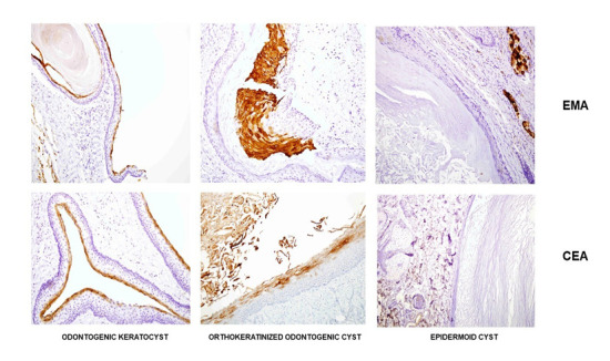
Materials and methods: An immunohistochemical analysis comprised of various cytokeratins, carcinoembryonic antigen (CEA), epithelial membrane antigen (EMA), epithelial cell adhesion molecules family member BerEP4, apoptosis-related markers bcl-2 and calretinin, stem cell marker CD44, tumor suppressor gene p63 and proliferation activity marker Ki-67 was performed on 25 OKCs, 8 OOCs and 11 ECs.
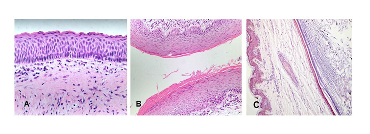
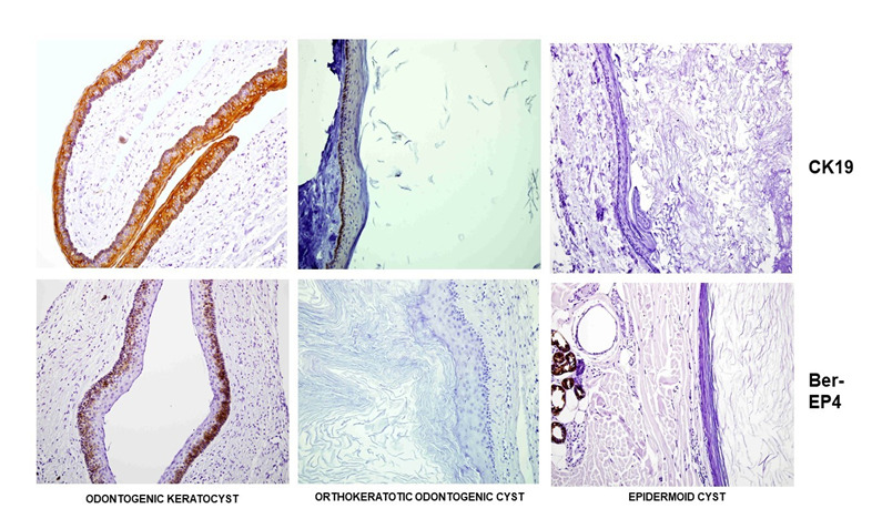
Results: CK5/6 was positive in all layers of both OKCs and OOCs, but only in the basal layer of all ECs. CK8/18 and CK19 revealed strong basal and suprabasal positivity in all OKCs, weak basal positivity in OOCs, and negative expression in all ECs. BerEP4 and Bcl-2 revealed positivity in all OKCs while being negative in OOCs and ECs.
Conclusions: The results of our study suggest that BerEP4 and Bcl-2 positivity may be useful in distinguishing between OKCs and the other keratinized jaw cysts. Orthokeratinized lesions within the jaw should be defined as OOCs, while intraosseal EC should be diagnosed only if immunohistochemical staining points to ectodermal origin, thus suggesting congenital or post-traumatic inclusion of the oral epithelium.
期刊介绍:
The Acta Stomatologica Croatica (ASCRO) is a leading scientific non-profit journal in the field of dental, oral and cranio-facial sciences during the past 44 years in Croatia. ASCRO publishes original scientific and clinical papers, preliminary communications, case reports, book reviews, letters to the editor and news. Review articles are published by invitation from the Editor-in-Chief by acclaimed professionals in distinct fields of dental medicine. All manuscripts are subjected to peer review process.

 求助内容:
求助内容: 应助结果提醒方式:


