Rapid cleavage of 6-[18F]fluoronicotinic acid prosthetic group governs BT12 glioblastoma xenograft uptake: implications for radiolabeling design of biomolecules
Pyry Dillemuth, Abiodun Ayo, Xiaoqing Zhuang, Petter Lövdahl, Heidi Liljenbäck, Salli Kärnä, Tatsiana Auchynnikava, Jonne Kunnas, Jesse Ponkamo, Maxwell W. G. Miner, Johan Rajander, Jessica M. Rosenholm, Anne Roivainen, Anu J. Airaksinen, Pirjo Laakkonen, Xiang-Guo Li
{"title":"Rapid cleavage of 6-[18F]fluoronicotinic acid prosthetic group governs BT12 glioblastoma xenograft uptake: implications for radiolabeling design of biomolecules","authors":"Pyry Dillemuth, Abiodun Ayo, Xiaoqing Zhuang, Petter Lövdahl, Heidi Liljenbäck, Salli Kärnä, Tatsiana Auchynnikava, Jonne Kunnas, Jesse Ponkamo, Maxwell W. G. Miner, Johan Rajander, Jessica M. Rosenholm, Anne Roivainen, Anu J. Airaksinen, Pirjo Laakkonen, Xiang-Guo Li","doi":"10.1186/s41181-025-00368-1","DOIUrl":null,"url":null,"abstract":"<div><h3>Background</h3><p>Peptides radiolabeled with fluorine-18 are frequently synthesized using prosthetic groups. Among them, activated esters of 6-[<sup>18</sup>F]fluoronicotinic acid ([<sup>18</sup>F]FNA) have been prepared and successfully employed for <sup>18</sup>F-labeling of diverse biomolecules, including peptides. The utility of [<sup>18</sup>F]FNA as a prosthetic compound has been demonstrated in both preclinical and clinical settings, including radiopharmaceuticals targeting prostate-specific membrane antigen and poly(ADP ribose) polymerase inhibitors. This study aims to evaluate a [<sup>18</sup>F]FNA-conjugated nonapeptide, [<sup>18</sup>F]FNA-<i>N</i>-CooP, for positron emission tomography imaging of intracranial BT12 glioblastoma xenografts in a mouse model. Additionally, this study highlights the importance of including control experiments with prosthetic compound alone when it constitutes a major radiometabolite.</p><h3>Results</h3><p>[<sup>18</sup>F]FNA-<i>N</i>-CooP successfully delineated intracranial glioblastoma xenografts yielding a standardized uptake value of 0.21 ± 0.03 (<i>n</i> = 4) and a tumor-to-brain ratio of 1.84 ± 0.29. Ex vivo autoradiography of tumor tissue showed a partial co-localization between radioactivity uptake and the target fatty acid binding protein 3 expression. However, in vivo instability of [<sup>18</sup>F]FNA-<i>N</i>-CooP was observed, with [<sup>18</sup>F]FNA identified as a major radiometabolite. Notably, control studies using [<sup>18</sup>F]FNA alone also visualized tumors, producing a standardized uptake value of 0.90 ± 0.10 (<i>n</i> = 4) and a tumor-to-brain ratio of 1.51 ± 0.08.</p><h3>Conclusions</h3><p>Both [<sup>18</sup>F]FNA-<i>N</i>-CooP and [<sup>18</sup>F]FNA enabled PET visualization of human glioblastoma in the mouse model. However, the prominent presence of [<sup>18</sup>F]FNA as radiometabolite complicates the interpretation of [<sup>18</sup>F]FNA-<i>N</i>-CooP PET data, suggesting that the observed radioactivity uptake may primarily originate from [<sup>18</sup>F]FNA and other radiometabolites. Enhancing peptide stability is essential for improving imaging specificity. This study underscores the critical need to assess the imaging contributions of prosthetic groups when they function as significant radiometabolites.</p></div>","PeriodicalId":534,"journal":{"name":"EJNMMI Radiopharmacy and Chemistry","volume":"10 1","pages":""},"PeriodicalIF":4.4000,"publicationDate":"2025-07-08","publicationTypes":"Journal Article","fieldsOfStudy":null,"isOpenAccess":false,"openAccessPdf":"https://www.ncbi.nlm.nih.gov/pmc/articles/PMC12238435/pdf/","citationCount":"0","resultStr":null,"platform":"Semanticscholar","paperid":null,"PeriodicalName":"EJNMMI Radiopharmacy and Chemistry","FirstCategoryId":"1085","ListUrlMain":"https://link.springer.com/article/10.1186/s41181-025-00368-1","RegionNum":0,"RegionCategory":null,"ArticlePicture":[],"TitleCN":null,"AbstractTextCN":null,"PMCID":null,"EPubDate":"","PubModel":"","JCR":"Q1","JCRName":"CHEMISTRY, INORGANIC & NUCLEAR","Score":null,"Total":0}
引用次数: 0
Abstract
Background
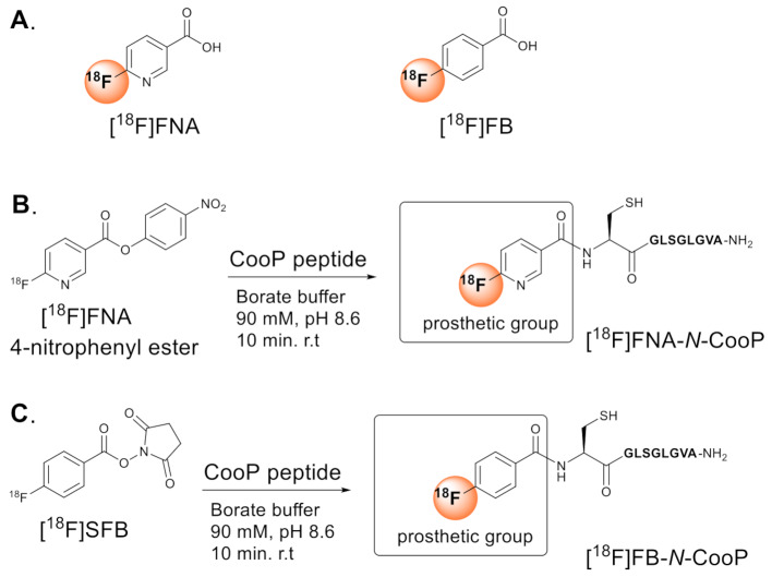
Peptides radiolabeled with fluorine-18 are frequently synthesized using prosthetic groups. Among them, activated esters of 6-[18F]fluoronicotinic acid ([18F]FNA) have been prepared and successfully employed for 18F-labeling of diverse biomolecules, including peptides. The utility of [18F]FNA as a prosthetic compound has been demonstrated in both preclinical and clinical settings, including radiopharmaceuticals targeting prostate-specific membrane antigen and poly(ADP ribose) polymerase inhibitors. This study aims to evaluate a [18F]FNA-conjugated nonapeptide, [18F]FNA-N-CooP, for positron emission tomography imaging of intracranial BT12 glioblastoma xenografts in a mouse model. Additionally, this study highlights the importance of including control experiments with prosthetic compound alone when it constitutes a major radiometabolite.
Results
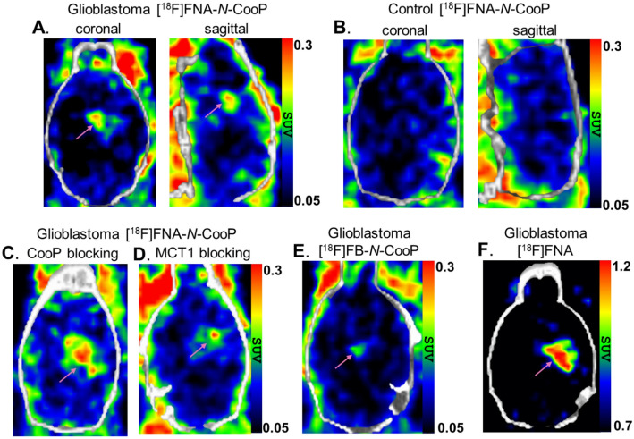
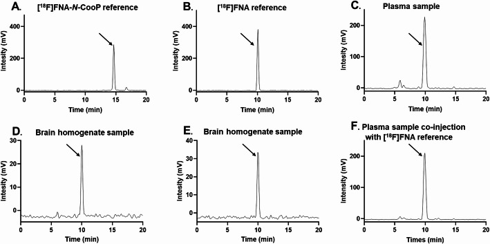
[18F]FNA-N-CooP successfully delineated intracranial glioblastoma xenografts yielding a standardized uptake value of 0.21 ± 0.03 (n = 4) and a tumor-to-brain ratio of 1.84 ± 0.29. Ex vivo autoradiography of tumor tissue showed a partial co-localization between radioactivity uptake and the target fatty acid binding protein 3 expression. However, in vivo instability of [18F]FNA-N-CooP was observed, with [18F]FNA identified as a major radiometabolite. Notably, control studies using [18F]FNA alone also visualized tumors, producing a standardized uptake value of 0.90 ± 0.10 (n = 4) and a tumor-to-brain ratio of 1.51 ± 0.08.
Conclusions
Both [18F]FNA-N-CooP and [18F]FNA enabled PET visualization of human glioblastoma in the mouse model. However, the prominent presence of [18F]FNA as radiometabolite complicates the interpretation of [18F]FNA-N-CooP PET data, suggesting that the observed radioactivity uptake may primarily originate from [18F]FNA and other radiometabolites. Enhancing peptide stability is essential for improving imaging specificity. This study underscores the critical need to assess the imaging contributions of prosthetic groups when they function as significant radiometabolites.

 求助内容:
求助内容: 应助结果提醒方式:


