Jacob Owens, Gunnar Whealy, Harvey Sekhon, Rustain Morgan, Craig Johnson, Lei Yu
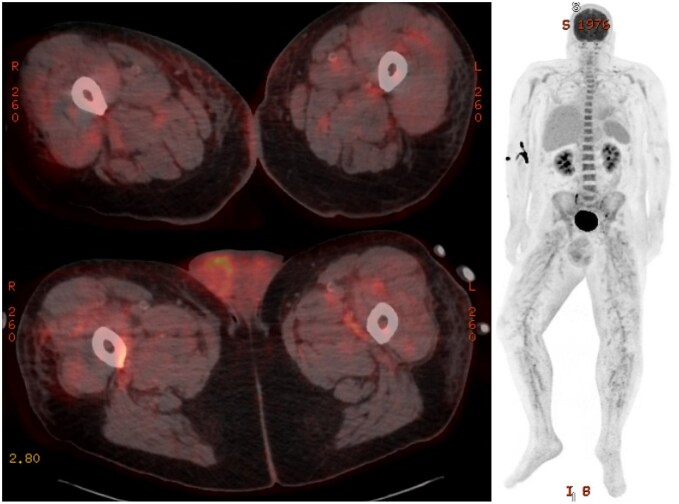
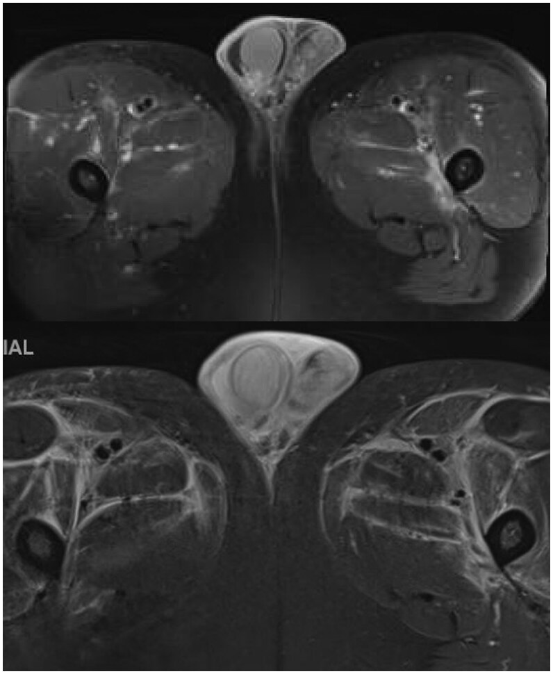
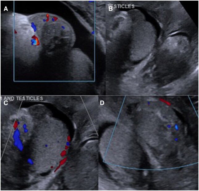
{"title":"Polyarteritis nodosa with testicular involvement: a rare case report highlighting the role of nuclear imaging and angiography in diagnosis.","authors":"Jacob Owens, Gunnar Whealy, Harvey Sekhon, Rustain Morgan, Craig Johnson, Lei Yu","doi":"10.1093/bjrcr/uaaf032","DOIUrl":null,"url":null,"abstract":"<p><p>Polyarteritis nodosa (PAN) is a systemic small to medium vessel vasculitis. It is often associated with hepatitis B infection and classically presents with cutaneous, gastrointestinal, or nervous system involvement. We present a case of a 56-year-old male who presented with a chief complaint of painful scrotal swelling. Initial ultrasound demonstrated concern for epididymitis, and the patient was started on appropriate antibiotics without improvement of symptoms, resulting in admission. Due to continued scrotal pain, fevers, and negative infectious work-up, F-18 fluorodeoxyglucose (FDG) PET/CT was obtained, revealing diffuse hypermetabolic activity throughout the medium to small arterial vasculature, concerning for vasculitis. Abdominopelvic angiography confirmed the diagnosis, and the patient was started on steroids with plans to initiate cyclophosphamide. Clinical testicular involvement is a rare presentation of PAN, although it is often seen at autopsy. Previously reported cases have presented with similar scrotal pain and tenderness in addition to constitutional symptoms, as well as treatment with steroids and immunosuppressive agents. While biopsy with histopathology or angiography often serves as the gold standard for the diagnosis of PAN, this case also demonstrates the diagnostic utility of nuclear medicine with F-18 FDG PET/CT. Polyarteritis nodosa typically demonstrates hypermetabolic activity of the small- to medium-sized vasculature on F-18 FDG PET/CT, most often in the lower extremities. With similar findings, this case contributes to reports that show the utility of nuclear imaging in diagnosing vasculitides.</p>","PeriodicalId":45216,"journal":{"name":"BJR Case Reports","volume":"11 4","pages":"uaaf032"},"PeriodicalIF":0.5000,"publicationDate":"2025-07-01","publicationTypes":"Journal Article","fieldsOfStudy":null,"isOpenAccess":false,"openAccessPdf":"https://www.ncbi.nlm.nih.gov/pmc/articles/PMC12228961/pdf/","citationCount":"0","resultStr":null,"platform":"Semanticscholar","paperid":null,"PeriodicalName":"BJR Case Reports","FirstCategoryId":"1085","ListUrlMain":"https://doi.org/10.1093/bjrcr/uaaf032","RegionNum":0,"RegionCategory":null,"ArticlePicture":[],"TitleCN":null,"AbstractTextCN":null,"PMCID":null,"EPubDate":"","PubModel":"","JCR":"Q4","JCRName":"RADIOLOGY, NUCLEAR MEDICINE & MEDICAL IMAGING","Score":null,"Total":0}
引用次数: 0
Abstract
Polyarteritis nodosa (PAN) is a systemic small to medium vessel vasculitis. It is often associated with hepatitis B infection and classically presents with cutaneous, gastrointestinal, or nervous system involvement. We present a case of a 56-year-old male who presented with a chief complaint of painful scrotal swelling. Initial ultrasound demonstrated concern for epididymitis, and the patient was started on appropriate antibiotics without improvement of symptoms, resulting in admission. Due to continued scrotal pain, fevers, and negative infectious work-up, F-18 fluorodeoxyglucose (FDG) PET/CT was obtained, revealing diffuse hypermetabolic activity throughout the medium to small arterial vasculature, concerning for vasculitis. Abdominopelvic angiography confirmed the diagnosis, and the patient was started on steroids with plans to initiate cyclophosphamide. Clinical testicular involvement is a rare presentation of PAN, although it is often seen at autopsy. Previously reported cases have presented with similar scrotal pain and tenderness in addition to constitutional symptoms, as well as treatment with steroids and immunosuppressive agents. While biopsy with histopathology or angiography often serves as the gold standard for the diagnosis of PAN, this case also demonstrates the diagnostic utility of nuclear medicine with F-18 FDG PET/CT. Polyarteritis nodosa typically demonstrates hypermetabolic activity of the small- to medium-sized vasculature on F-18 FDG PET/CT, most often in the lower extremities. With similar findings, this case contributes to reports that show the utility of nuclear imaging in diagnosing vasculitides.

 求助内容:
求助内容: 应助结果提醒方式:


