Mohammed Obaid, Arman Rahmim, William P Segars, Julia Brosch-Lenz, Carlos Uribe
{"title":"S-values for bone marrow dosimetry in preclinical radiopharmaceutical studies with rodents.","authors":"Mohammed Obaid, Arman Rahmim, William P Segars, Julia Brosch-Lenz, Carlos Uribe","doi":"10.1186/s40658-025-00752-5","DOIUrl":null,"url":null,"abstract":"<p><strong>Background: </strong>Development of novel radiopharmaceuticals involves dosimetry calculations to validate safety and aid with selection of those that should be translated into the clinical environment. Dosimetry is critical for limiting radiation damage to organs at risk. The bone marrow is a limiting organ in radiopharmaceutical therapies (RPTs) for metastatic prostate cancer, for example, but there is room for improvement of bone marrow dosimetry in preclinical studies. Bone marrow S-values for Lutetium-177 (<sup>177</sup>Lu) in rodents have been published but they have not included tumor xenografts inoculated in the shoulder, which is how radiopharmaceuticals are often tested. Here, we aim at performing Monte Carlo simulations on digital mice phantoms including tumor xenografts, and to determine new bone marrow S-values that can potentially improve our understanding of the effect of RPTs in blood cells.</p><p><strong>Methods: </strong>S-values for <sup>177</sup>Lu were simulated in the 4D Mouse Whole Body (MOBY) phantom, a hybrid voxel-based mouse model, using GATE v9.3 MC toolkit. Two phantoms of different resolutions and equal mass were created. 3D dose distributions were simulated and the corresponding organ to organ S-values were calculated. The resulting S-values were validated against reference values from OLINDA v2.2.3. Later, tumours of varying sizes were placed in the left shoulder and tumour-to-organ S-values were calculated from MC simulations with a <sup>177</sup>Lu source placed uniformly in these tumours.</p><p><strong>Results: </strong>The phantoms simulated here differed from the OLINDA phantom in both organ mass and geometry for many tissues; S-value deviations from OLINDA were correlated with these differences, as reported in previous studies, and ranged from 2% for the kidney self-dose in the higher resolution (HR) phantom to 477% for S(skeleton←spleen) in the lower resolution (LR) phantom. S-values were simulated for the bone marrow in both phantoms; cross-dose values were greatest from the skeleton, brain, and lungs, while cross-doses from the simulated tumours were approximately constant at 3 × 10<sup>-15</sup> Gy Bq<sup>-1</sup> s<sup>-1</sup> across all tumour sizes. The components of the skeleton receiving the greatest tumour cross-doses from the tumours were the spine, skull and marrow. S-values targeting the bone marrow were compared to similar values from a previous study, whose phantom differed in tissue composition-discrepancies ranged from 6% for S(BM←kidneys) at LR to 87% for S(BM←BM) at HR. In general, relative uncertainty in dose and dose factor deposited from one tissue to another was inversely proportional to the corresponding S-value magnitude, and lower uncertainties were yielded from simulations in the LR, large-voxel phantom.</p><p><strong>Conclusion: </strong>Using the MOBY digital mouse phantom, we simulated bone marrow S-values for <sup>177</sup>Lu. We hope these values help researchers perform preclinical dosimetry in rodents including bone marrow and tumor xenografts and facilitate the translation of novel radiopharmaceuticals.</p>","PeriodicalId":11559,"journal":{"name":"EJNMMI Physics","volume":"12 1","pages":"67"},"PeriodicalIF":3.2000,"publicationDate":"2025-07-08","publicationTypes":"Journal Article","fieldsOfStudy":null,"isOpenAccess":false,"openAccessPdf":"https://www.ncbi.nlm.nih.gov/pmc/articles/PMC12238459/pdf/","citationCount":"0","resultStr":null,"platform":"Semanticscholar","paperid":null,"PeriodicalName":"EJNMMI Physics","FirstCategoryId":"3","ListUrlMain":"https://doi.org/10.1186/s40658-025-00752-5","RegionNum":2,"RegionCategory":"医学","ArticlePicture":[],"TitleCN":null,"AbstractTextCN":null,"PMCID":null,"EPubDate":"","PubModel":"","JCR":"Q2","JCRName":"RADIOLOGY, NUCLEAR MEDICINE & MEDICAL IMAGING","Score":null,"Total":0}
引用次数: 0
Abstract
Background: Development of novel radiopharmaceuticals involves dosimetry calculations to validate safety and aid with selection of those that should be translated into the clinical environment. Dosimetry is critical for limiting radiation damage to organs at risk. The bone marrow is a limiting organ in radiopharmaceutical therapies (RPTs) for metastatic prostate cancer, for example, but there is room for improvement of bone marrow dosimetry in preclinical studies. Bone marrow S-values for Lutetium-177 (177Lu) in rodents have been published but they have not included tumor xenografts inoculated in the shoulder, which is how radiopharmaceuticals are often tested. Here, we aim at performing Monte Carlo simulations on digital mice phantoms including tumor xenografts, and to determine new bone marrow S-values that can potentially improve our understanding of the effect of RPTs in blood cells.
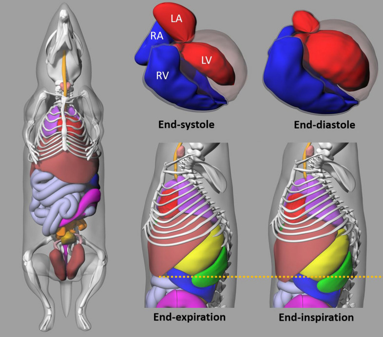
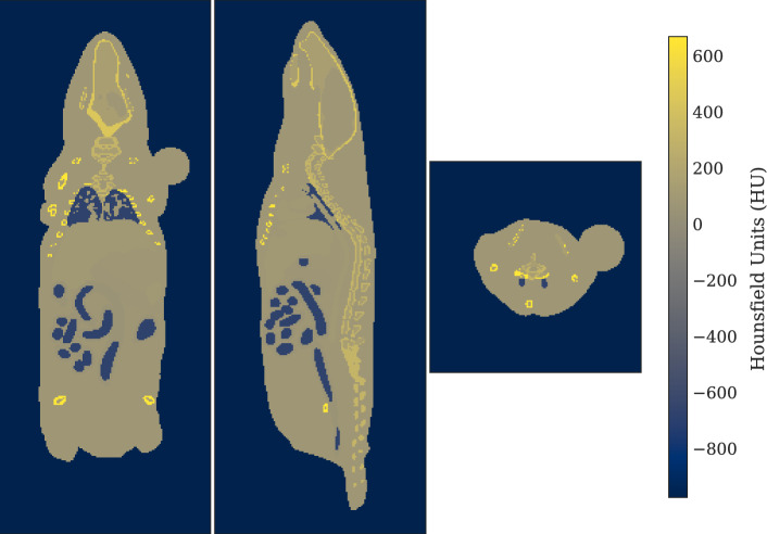
Methods: S-values for 177Lu were simulated in the 4D Mouse Whole Body (MOBY) phantom, a hybrid voxel-based mouse model, using GATE v9.3 MC toolkit. Two phantoms of different resolutions and equal mass were created. 3D dose distributions were simulated and the corresponding organ to organ S-values were calculated. The resulting S-values were validated against reference values from OLINDA v2.2.3. Later, tumours of varying sizes were placed in the left shoulder and tumour-to-organ S-values were calculated from MC simulations with a 177Lu source placed uniformly in these tumours.
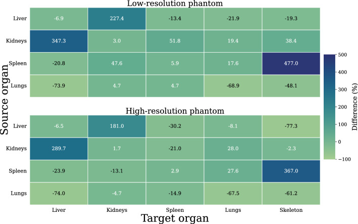
Results: The phantoms simulated here differed from the OLINDA phantom in both organ mass and geometry for many tissues; S-value deviations from OLINDA were correlated with these differences, as reported in previous studies, and ranged from 2% for the kidney self-dose in the higher resolution (HR) phantom to 477% for S(skeleton←spleen) in the lower resolution (LR) phantom. S-values were simulated for the bone marrow in both phantoms; cross-dose values were greatest from the skeleton, brain, and lungs, while cross-doses from the simulated tumours were approximately constant at 3 × 10-15 Gy Bq-1 s-1 across all tumour sizes. The components of the skeleton receiving the greatest tumour cross-doses from the tumours were the spine, skull and marrow. S-values targeting the bone marrow were compared to similar values from a previous study, whose phantom differed in tissue composition-discrepancies ranged from 6% for S(BM←kidneys) at LR to 87% for S(BM←BM) at HR. In general, relative uncertainty in dose and dose factor deposited from one tissue to another was inversely proportional to the corresponding S-value magnitude, and lower uncertainties were yielded from simulations in the LR, large-voxel phantom.
Conclusion: Using the MOBY digital mouse phantom, we simulated bone marrow S-values for 177Lu. We hope these values help researchers perform preclinical dosimetry in rodents including bone marrow and tumor xenografts and facilitate the translation of novel radiopharmaceuticals.
期刊介绍:
EJNMMI Physics is an international platform for scientists, users and adopters of nuclear medicine with a particular interest in physics matters. As a companion journal to the European Journal of Nuclear Medicine and Molecular Imaging, this journal has a multi-disciplinary approach and welcomes original materials and studies with a focus on applied physics and mathematics as well as imaging systems engineering and prototyping in nuclear medicine. This includes physics-driven approaches or algorithms supported by physics that foster early clinical adoption of nuclear medicine imaging and therapy.

 求助内容:
求助内容: 应助结果提醒方式:


