Automated volumetric estimation of six basic thoracic and abdominal organs in postmortem computed tomography data using deep learning techniques
IF 1
Q4 RADIOLOGY, NUCLEAR MEDICINE & MEDICAL IMAGING
引用次数: 0
Abstract
Purpose
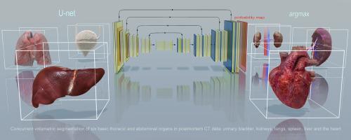
Computed tomography (CT) has become a widely adopted and standard procedure as an adjunct to autopsies in numerous countries. However, owing to the high number of cases and the limited availability of skilled practitioners, the need to streamline the diagnostic process has spurred the advancement of automated solutions. These solutions leverage deep learning methodologies to potentially automate diagnoses by analyzing postmortem CT data. Here, we show how deep learning techniques enable segmentation and volume evaluation to be concurrently performed for six basic thoracic and abdominal organs in postmortem CT data: the heart, lungs, liver, spleen, kidneys, and urinary bladder. Based on these automated volumetric estimations we automatically derived the weight of the heart, lungs, liver, spleen, and kidneys.
Methods
We developed a convolutional neural network tailored for conducting volumetric data segmentation in postmortem computed tomography images based on the U-Net architecture.
Results
Our best model achieved an overall Dice score (F1 score) of 0.907±0.029. The heart, lung, and liver yielded higher scores than did the spleen, kidneys, and urinary bladder. We also automated the weight calculation of the heart, lungs, liver, spleen, and kidneys.
Conclusion
Our study demonstrated that a convolution neural network such as U-Net could reliably estimate concurrently the volumes of six basic thoracic and abdominal organs from postmortem CT data. Our study also shows how this information can be subsequently used to automatically estimate their weight. However, post- and perimortem changes pose substantial challenges for automatically processing postmortem CT data.
使用深度学习技术对死后计算机断层扫描数据中六个基本胸腹器官的自动体积估计
在许多国家,计算机断层扫描(CT)已成为一种广泛采用的标准程序,作为尸检的辅助手段。然而,由于病例数量多,技术熟练的从业人员有限,简化诊断过程的需要促使了自动化解决方案的发展。这些解决方案利用深度学习方法,通过分析死后CT数据来自动诊断。在这里,我们展示了深度学习技术如何能够同时对死后CT数据中的六个基本胸部和腹部器官(心脏、肺、肝脏、脾脏、肾脏和膀胱)进行分割和体积评估。基于这些自动的体积估计,我们自动得出了心脏、肺、肝、脾和肾的重量。方法基于U-Net架构开发了一种卷积神经网络,用于对尸体计算机断层扫描图像进行体积数据分割。结果最佳模型的Dice总分(F1分)为0.907±0.029。心脏、肺和肝脏的得分高于脾脏、肾脏和膀胱。我们还自动计算了心脏、肺、肝脏、脾脏和肾脏的重量。结论基于U-Net的卷积神经网络可以可靠地从死后CT数据中同时估计六个基本胸腹器官的体积。我们的研究还显示了如何利用这些信息来自动估计他们的体重。然而,死后和死前的变化对自动处理死后CT数据提出了实质性的挑战。
本文章由计算机程序翻译,如有差异,请以英文原文为准。
求助全文
约1分钟内获得全文
求助全文
来源期刊
Forensic Imaging
RADIOLOGY, NUCLEAR MEDICINE & MEDICAL IMAGING-
CiteScore
2.20
自引率
27.30%
发文量
39
 求助内容:
求助内容: 应助结果提醒方式:


