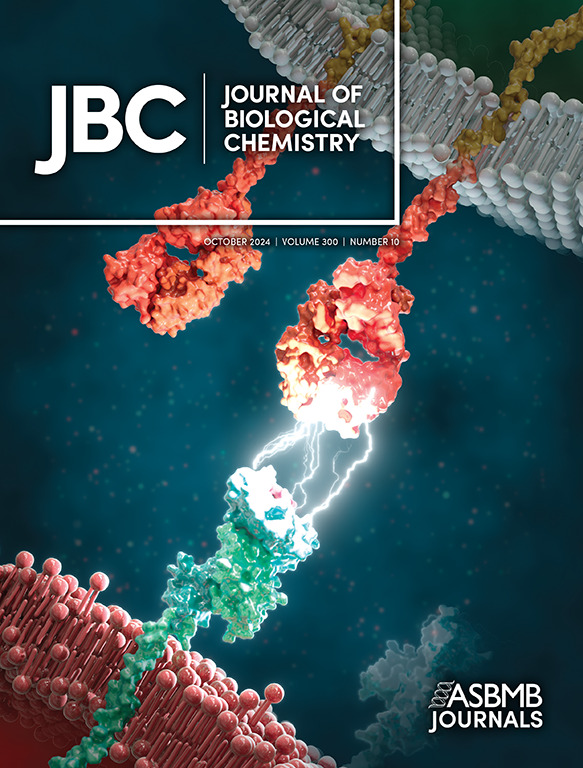
Inflamed endothelial cells express S1PR1 inhibitor CD69 to induce vascular leak.
IF 4
2区 生物学
Q2 BIOCHEMISTRY & MOLECULAR BIOLOGY
引用次数: 0
Abstract
Inflammation disrupts endothelial barrier function and causes vascular leak into the tissue parenchyma. Sphingosine 1-phosphate receptor-1 (S1PR1) in endothelial cells (EC) is a key inducer of endothelial junctions and barrier function. We report here that endothelial cell activation by the cytokine TNFα and TLR3 agonist poly-inosine/cytosine (pI:C) induces the lymphocyte activation molecule CD69 via the canonical NFκB pathway. EC CD69 stimulates endocytosis of S1PR1, inhibits its downstream intracellular signaling events and barrier function. Administration of TLR4 or TLR3 agonists or intranasal infection of mouse-adapted influenza virus (H1N1) or coronavirus (MHV-A59) induced CD69 in lung endothelial cells. AAV-mediated overexpression of CD69 in lung EC leads to decreased cell-surface expression of S1PR1 and tight junction protein Claudin-5, concomitantly with increased vascular permeability in the lungs. Further, lung vascular leak at the peak of H1N1 infection is attenuated in a genetic mouse model which lacks CD69 in the endothelium. These data suggest that endothelial activation during inflammation and viral host-defense induces CD69 which downregulates S1PR1 to induce vascular leakage. CD69 induction during endothelial dysfunction may drive exaggerated inflammation by antagonizing the endothelial protective S1PR1 pathway.炎症内皮细胞表达S1PR1抑制剂CD69诱导血管渗漏。
炎症破坏内皮屏障功能,导致血管渗漏到组织实质。内皮细胞(EC)中的鞘氨醇1-磷酸受体1 (S1PR1)是内皮细胞连接和屏障功能的关键诱导剂。我们在这里报道了内皮细胞被细胞因子TNFα和TLR3激动剂多肌苷/胞嘧啶(pI:C)激活,通过典型的NFκB途径诱导淋巴细胞激活分子CD69。EC CD69刺激S1PR1的内吞作用,抑制其下游胞内信号事件和屏障功能。给予TLR4或TLR3激动剂或鼻内感染小鼠适应性流感病毒(H1N1)或冠状病毒(MHV-A59)可诱导肺内皮细胞中的CD69。aav介导的CD69在肺EC中的过表达导致S1PR1和紧密连接蛋白Claudin-5的细胞表面表达降低,同时肺血管通透性增加。此外,在内皮中缺乏CD69的遗传小鼠模型中,H1N1感染高峰时的肺血管泄漏被减弱。这些数据表明,炎症和病毒宿主防御期间内皮细胞的激活可诱导CD69下调S1PR1,从而诱导血管渗漏。内皮功能障碍过程中CD69的诱导可能通过拮抗内皮保护性通路S1PR1而导致炎症加重。
本文章由计算机程序翻译,如有差异,请以英文原文为准。
求助全文
约1分钟内获得全文
求助全文
来源期刊

Journal of Biological Chemistry
Biochemistry, Genetics and Molecular Biology-Biochemistry
自引率
4.20%
发文量
1233
期刊介绍:
The Journal of Biological Chemistry welcomes high-quality science that seeks to elucidate the molecular and cellular basis of biological processes. Papers published in JBC can therefore fall under the umbrellas of not only biological chemistry, chemical biology, or biochemistry, but also allied disciplines such as biophysics, systems biology, RNA biology, immunology, microbiology, neurobiology, epigenetics, computational biology, ’omics, and many more. The outcome of our focus on papers that contribute novel and important mechanistic insights, rather than on a particular topic area, is that JBC is truly a melting pot for scientists across disciplines. In addition, JBC welcomes papers that describe methods that will help scientists push their biochemical inquiries forward and resources that will be of use to the research community.
 求助内容:
求助内容: 应助结果提醒方式:


