Using Segment Anything Model 2 for Zero-Shot 3D Segmentation of Abdominal Organs in Computed Tomography Scans to Adapt Video Tracking Capabilities for 3D Medical Imaging: Algorithm Development and Validation.
{"title":"Using Segment Anything Model 2 for Zero-Shot 3D Segmentation of Abdominal Organs in Computed Tomography Scans to Adapt Video Tracking Capabilities for 3D Medical Imaging: Algorithm Development and Validation.","authors":"Yosuke Yamagishi, Shouhei Hanaoka, Tomohiro Kikuchi, Takahiro Nakao, Yuta Nakamura, Yukihiro Nomura, Soichiro Miki, Takeharu Yoshikawa, Osamu Abe","doi":"10.2196/72109","DOIUrl":null,"url":null,"abstract":"<p><strong>Background: </strong>Medical image segmentation is crucial for diagnosis and treatment planning in radiology, but it traditionally requires extensive manual effort and specialized training data. With its novel video tracking capabilities, the Segment Anything Model 2 (SAM 2) presents a potential solution for automated 3D medical image segmentation without the need for domain-specific training. However, its effectiveness in medical applications, particularly in abdominal computed tomography (CT) imaging remains unexplored.</p><p><strong>Objective: </strong>The aim of this study was to evaluate the zero-shot performance of SAM 2 in 3D segmentation of abdominal organs in CT scans and to investigate the effects of prompt settings on segmentation results.</p><p><strong>Methods: </strong>In this retrospective study, we used a subset of the TotalSegmentator CT dataset from eight institutions to assess SAM 2's ability to segment eight abdominal organs. Segmentation was initiated from three different z-coordinate levels (caudal, mid, and cranial levels) of each organ. Performance was measured using the dice similarity coefficient (DSC). We also analyzed the impact of \"negative prompts,\" which explicitly exclude certain regions from the segmentation process, on accuracy.</p><p><strong>Results: </strong>A total of 123 patients (mean age 60.7, SD 15.5 years; 63 men, 60 women) were evaluated. As a zero-shot approach, larger organs with clear boundaries demonstrated high segmentation performance, with mean DSCs as follows: liver, 0.821 (SD 0.192); right kidney, 0.862 (SD 0.212); left kidney, 0.870 (SD 0.154); and spleen, 0.891 (SD 0.131). Smaller organs showed lower performance: gallbladder, 0.531 (SD 0.291); pancreas, 0.361 (SD 0.197); and adrenal glands-right, 0.203 (SD 0.222) and left, 0.308 (SD 0.234). The initial slice for segmentation and the use of negative prompts significantly influenced the results. By removing negative prompts from the input, the DSCs significantly decreased for six organs.</p><p><strong>Conclusions: </strong>SAM 2 demonstrated promising zero-shot performance in segmenting certain abdominal organs in CT scans, particularly larger organs. Performance was significantly influenced by input negative prompts and initial slice selection, highlighting the importance of optimizing these factors.</p>","PeriodicalId":73551,"journal":{"name":"JMIR AI","volume":"4 ","pages":"e72109"},"PeriodicalIF":2.0000,"publicationDate":"2025-04-29","publicationTypes":"Journal Article","fieldsOfStudy":null,"isOpenAccess":false,"openAccessPdf":"https://www.ncbi.nlm.nih.gov/pmc/articles/PMC12231515/pdf/","citationCount":"0","resultStr":null,"platform":"Semanticscholar","paperid":null,"PeriodicalName":"JMIR AI","FirstCategoryId":"1085","ListUrlMain":"https://doi.org/10.2196/72109","RegionNum":0,"RegionCategory":null,"ArticlePicture":[],"TitleCN":null,"AbstractTextCN":null,"PMCID":null,"EPubDate":"","PubModel":"","JCR":"","JCRName":"","Score":null,"Total":0}
引用次数: 0
Abstract
Background: Medical image segmentation is crucial for diagnosis and treatment planning in radiology, but it traditionally requires extensive manual effort and specialized training data. With its novel video tracking capabilities, the Segment Anything Model 2 (SAM 2) presents a potential solution for automated 3D medical image segmentation without the need for domain-specific training. However, its effectiveness in medical applications, particularly in abdominal computed tomography (CT) imaging remains unexplored.
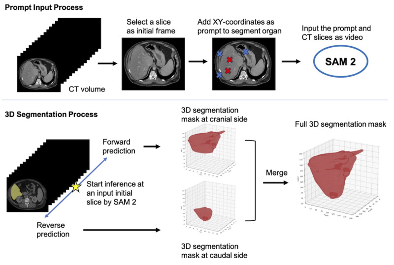
Objective: The aim of this study was to evaluate the zero-shot performance of SAM 2 in 3D segmentation of abdominal organs in CT scans and to investigate the effects of prompt settings on segmentation results.
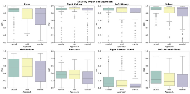
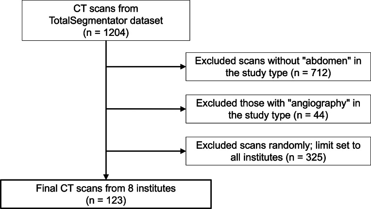
Methods: In this retrospective study, we used a subset of the TotalSegmentator CT dataset from eight institutions to assess SAM 2's ability to segment eight abdominal organs. Segmentation was initiated from three different z-coordinate levels (caudal, mid, and cranial levels) of each organ. Performance was measured using the dice similarity coefficient (DSC). We also analyzed the impact of "negative prompts," which explicitly exclude certain regions from the segmentation process, on accuracy.
Results: A total of 123 patients (mean age 60.7, SD 15.5 years; 63 men, 60 women) were evaluated. As a zero-shot approach, larger organs with clear boundaries demonstrated high segmentation performance, with mean DSCs as follows: liver, 0.821 (SD 0.192); right kidney, 0.862 (SD 0.212); left kidney, 0.870 (SD 0.154); and spleen, 0.891 (SD 0.131). Smaller organs showed lower performance: gallbladder, 0.531 (SD 0.291); pancreas, 0.361 (SD 0.197); and adrenal glands-right, 0.203 (SD 0.222) and left, 0.308 (SD 0.234). The initial slice for segmentation and the use of negative prompts significantly influenced the results. By removing negative prompts from the input, the DSCs significantly decreased for six organs.
Conclusions: SAM 2 demonstrated promising zero-shot performance in segmenting certain abdominal organs in CT scans, particularly larger organs. Performance was significantly influenced by input negative prompts and initial slice selection, highlighting the importance of optimizing these factors.

 求助内容:
求助内容: 应助结果提醒方式:


