Laurien De Roeck, Rob Colaes, Patrick Dupont, Stefan Sunaert, Steven De Vleeschouwer, Paul M Clement, Charlotte Sleurs, Maarten Lambrecht
{"title":"Increased functional network segregation in glioma patients posttherapy: A neurological compensatory response or catastrophe for cognition?","authors":"Laurien De Roeck, Rob Colaes, Patrick Dupont, Stefan Sunaert, Steven De Vleeschouwer, Paul M Clement, Charlotte Sleurs, Maarten Lambrecht","doi":"10.1162/netn_a_00449","DOIUrl":null,"url":null,"abstract":"<p><p>The brain operates through networks of interconnected regions, which can be disrupted by glial tumors and their treatment. This study investigates associations between this altered functional network topology and cognition in gliomas. We studied 50 adult glioma survivors (>1-year posttherapy) and 50 healthy controls. Participants underwent cognitive assessments across six domains and an 8-min resting-state functional MRI. Based on the BOLD signal, partial correlations were computed among 78 brain regions. From their absolute values, whole-brain and nodal graph metrics were derived and normalized to random graphs. Group differences in whole-brain and nodal graph metrics were assessed with Mann-Whitney <i>U</i> tests and mixed-design analyses of variance, respectively. Metrics exhibiting significant intergroup differences were correlated with cognitive scores, with <i>p</i> <sub>bonf</sub> < 0.050 indicating significance. Among controls, 8 of 78 nodes were identified as hubs. Patients exhibited significantly higher whole-brain clustering, correlating with intelligence (<i>r</i>(98) = -0.409, <i>p</i> <sub>bonf</sub> < 0.001) and executive functioning (<i>r</i>(98) = 0.300, <i>p</i> <sub>bonf</sub> = 0.014). Lower centrality, higher nodal clustering, and assortativity were also observed in patients, particularly in hubs, correlating with language and executive functioning, respectively (all <i>r</i>(98) > 0.300, <i>p</i> <sub>bonf</sub> < 0.050). Glioma patients commonly experience cognitive deficits alongside posttreatment alterations in functional network topology. Alterations in clustering, assortativity, and centrality may specifically act as compensatory mechanisms, significantly influencing cognitive functioning.</p>","PeriodicalId":48520,"journal":{"name":"Network Neuroscience","volume":"9 2","pages":"743-760"},"PeriodicalIF":3.1000,"publicationDate":"2025-06-27","publicationTypes":"Journal Article","fieldsOfStudy":null,"isOpenAccess":false,"openAccessPdf":"https://www.ncbi.nlm.nih.gov/pmc/articles/PMC12226146/pdf/","citationCount":"0","resultStr":null,"platform":"Semanticscholar","paperid":null,"PeriodicalName":"Network Neuroscience","FirstCategoryId":"3","ListUrlMain":"https://doi.org/10.1162/netn_a_00449","RegionNum":3,"RegionCategory":"医学","ArticlePicture":[],"TitleCN":null,"AbstractTextCN":null,"PMCID":null,"EPubDate":"2025/1/1 0:00:00","PubModel":"eCollection","JCR":"Q2","JCRName":"NEUROSCIENCES","Score":null,"Total":0}
引用次数: 0
Abstract
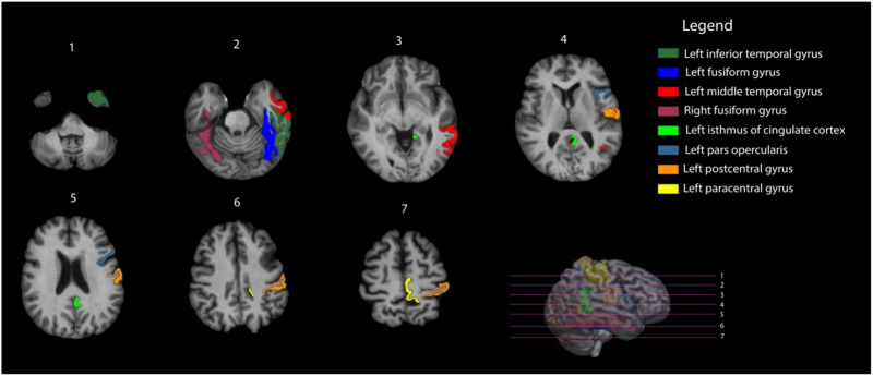
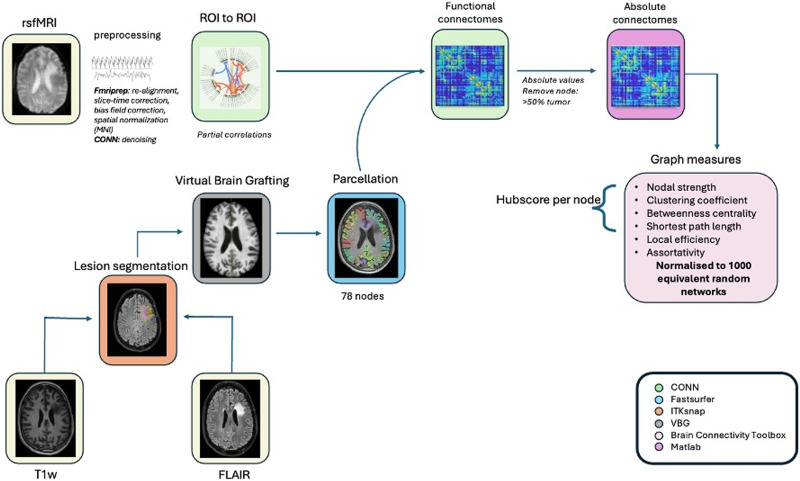
The brain operates through networks of interconnected regions, which can be disrupted by glial tumors and their treatment. This study investigates associations between this altered functional network topology and cognition in gliomas. We studied 50 adult glioma survivors (>1-year posttherapy) and 50 healthy controls. Participants underwent cognitive assessments across six domains and an 8-min resting-state functional MRI. Based on the BOLD signal, partial correlations were computed among 78 brain regions. From their absolute values, whole-brain and nodal graph metrics were derived and normalized to random graphs. Group differences in whole-brain and nodal graph metrics were assessed with Mann-Whitney U tests and mixed-design analyses of variance, respectively. Metrics exhibiting significant intergroup differences were correlated with cognitive scores, with pbonf < 0.050 indicating significance. Among controls, 8 of 78 nodes were identified as hubs. Patients exhibited significantly higher whole-brain clustering, correlating with intelligence (r(98) = -0.409, pbonf < 0.001) and executive functioning (r(98) = 0.300, pbonf = 0.014). Lower centrality, higher nodal clustering, and assortativity were also observed in patients, particularly in hubs, correlating with language and executive functioning, respectively (all r(98) > 0.300, pbonf < 0.050). Glioma patients commonly experience cognitive deficits alongside posttreatment alterations in functional network topology. Alterations in clustering, assortativity, and centrality may specifically act as compensatory mechanisms, significantly influencing cognitive functioning.


 求助内容:
求助内容: 应助结果提醒方式:


