Mahmudur Rahman, Jifan Gao, Kyle A Carey, Dana P Edelson, Askar Afshar, John W Garrett, Guanhua Chen, Majid Afshar, Matthew M Churpek
{"title":"Comparison of Deep Learning Approaches Using Chest Radiographs for Predicting Clinical Deterioration: Retrospective Observational Study.","authors":"Mahmudur Rahman, Jifan Gao, Kyle A Carey, Dana P Edelson, Askar Afshar, John W Garrett, Guanhua Chen, Majid Afshar, Matthew M Churpek","doi":"10.2196/67144","DOIUrl":null,"url":null,"abstract":"<p><strong>Background: </strong>The early detection of clinical deterioration and timely intervention for hospitalized patients can improve patient outcomes. The currently existing early warning systems rely on variables from structured data, such as vital signs and laboratory values, and do not incorporate other potentially predictive data modalities. Because respiratory failure is a common cause of deterioration, chest radiographs are often acquired in patients with clinical deterioration, which may be informative for predicting their risk of intensive care unit (ICU) transfer.</p><p><strong>Objective: </strong>This study aimed to compare and validate different computer vision models and data augmentation approaches with chest radiographs for predicting clinical deterioration.</p><p><strong>Methods: </strong>This retrospective observational study included adult patients hospitalized at the University of Wisconsin Health System between 2009 and 2020 with an elevated electronic cardiac arrest risk triage (eCART) score, a validated clinical deterioration early warning score, on the medical-surgical wards. Patients with a chest radiograph obtained within 48 hours prior to the elevated score were included in this study. Five computer vision model architectures (VGG16, DenseNet121, Vision Transformer, ResNet50, and Inception V3) and four data augmentation methods (histogram normalization, random flip, random Gaussian noise, and random rotate) were compared using the area under the receiver operating characteristic curve (AUROC) and the area under the precision-recall curve (AUPRC) for predicting clinical deterioration (ie, ICU transfer or ward death in the following 24 hours).</p><p><strong>Results: </strong>The study included 21,817 patient admissions, of which 1655 (7.6%) experienced clinical deterioration. The DenseNet121 model pretrained on chest radiograph datasets with histogram normalization and random Gaussian noise augmentation had the highest discrimination (AUROC 0.734 and AUPRC 0.414), while the vision transformer having 24 transformer blocks with random rotate augmentation had the lowest discrimination (AUROC 0.598).</p><p><strong>Conclusions: </strong>The study shows the potential of chest radiographs in deep learning models for predicting clinical deterioration. The DenseNet121 architecture pretrained with chest radiographs performed better than other architectures in most experiments, and the addition of histogram normalization with random Gaussian noise data augmentation may enhance the performance of DenseNet121 and pretrained VGG16 architectures.</p>","PeriodicalId":73551,"journal":{"name":"JMIR AI","volume":"4 ","pages":"e67144"},"PeriodicalIF":2.0000,"publicationDate":"2025-04-10","publicationTypes":"Journal Article","fieldsOfStudy":null,"isOpenAccess":false,"openAccessPdf":"https://www.ncbi.nlm.nih.gov/pmc/articles/PMC12223691/pdf/","citationCount":"0","resultStr":null,"platform":"Semanticscholar","paperid":null,"PeriodicalName":"JMIR AI","FirstCategoryId":"1085","ListUrlMain":"https://doi.org/10.2196/67144","RegionNum":0,"RegionCategory":null,"ArticlePicture":[],"TitleCN":null,"AbstractTextCN":null,"PMCID":null,"EPubDate":"","PubModel":"","JCR":"","JCRName":"","Score":null,"Total":0}
引用次数: 0
Abstract
Background: The early detection of clinical deterioration and timely intervention for hospitalized patients can improve patient outcomes. The currently existing early warning systems rely on variables from structured data, such as vital signs and laboratory values, and do not incorporate other potentially predictive data modalities. Because respiratory failure is a common cause of deterioration, chest radiographs are often acquired in patients with clinical deterioration, which may be informative for predicting their risk of intensive care unit (ICU) transfer.
Objective: This study aimed to compare and validate different computer vision models and data augmentation approaches with chest radiographs for predicting clinical deterioration.
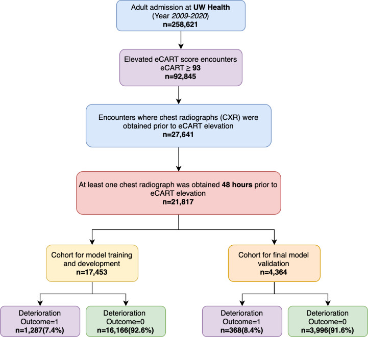
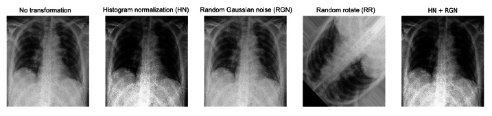
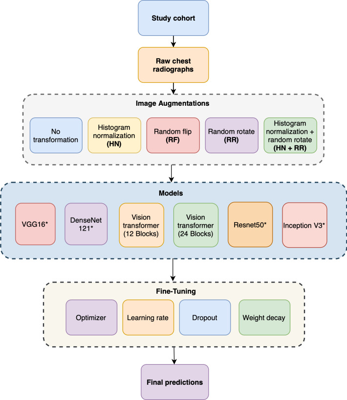
Methods: This retrospective observational study included adult patients hospitalized at the University of Wisconsin Health System between 2009 and 2020 with an elevated electronic cardiac arrest risk triage (eCART) score, a validated clinical deterioration early warning score, on the medical-surgical wards. Patients with a chest radiograph obtained within 48 hours prior to the elevated score were included in this study. Five computer vision model architectures (VGG16, DenseNet121, Vision Transformer, ResNet50, and Inception V3) and four data augmentation methods (histogram normalization, random flip, random Gaussian noise, and random rotate) were compared using the area under the receiver operating characteristic curve (AUROC) and the area under the precision-recall curve (AUPRC) for predicting clinical deterioration (ie, ICU transfer or ward death in the following 24 hours).
Results: The study included 21,817 patient admissions, of which 1655 (7.6%) experienced clinical deterioration. The DenseNet121 model pretrained on chest radiograph datasets with histogram normalization and random Gaussian noise augmentation had the highest discrimination (AUROC 0.734 and AUPRC 0.414), while the vision transformer having 24 transformer blocks with random rotate augmentation had the lowest discrimination (AUROC 0.598).
Conclusions: The study shows the potential of chest radiographs in deep learning models for predicting clinical deterioration. The DenseNet121 architecture pretrained with chest radiographs performed better than other architectures in most experiments, and the addition of histogram normalization with random Gaussian noise data augmentation may enhance the performance of DenseNet121 and pretrained VGG16 architectures.

 求助内容:
求助内容: 应助结果提醒方式:


