Bartolomeo Cassano, Ludovica Miseo, Sara Ungania, Marco D'Andrea, Federica Murtas, Massimiliano Pacilio, Marta Bottero, Daria Maccora, Rosa Sciuto, Giulio Eugenio Vallati, Antonella Soriani, Giuseppe Iaccarino
{"title":"Dosimetric study on radioembolization with 166Ho poly L-lactic acid microspheres: dead time effects on prediction power.","authors":"Bartolomeo Cassano, Ludovica Miseo, Sara Ungania, Marco D'Andrea, Federica Murtas, Massimiliano Pacilio, Marta Bottero, Daria Maccora, Rosa Sciuto, Giulio Eugenio Vallati, Antonella Soriani, Giuseppe Iaccarino","doi":"10.1186/s40658-025-00779-8","DOIUrl":null,"url":null,"abstract":"<p><strong>Background: </strong><sup>166</sup>Ho-poly-L-lactic acid microspheres (<sup>166</sup>Ho-PLLA) offer the advantage of using the same microspheres for both Scout and Therapeutic Administrations (SA and TA) in radioembolization compared to <sup>90</sup>Y. This study aimed to quantify and correct dead time (DT) effects in dose estimation and assess the predictive power of SA on TA.</p><p><strong>Methods: </strong>A 1.9 GBq <sup>166</sup>Ho-PLLA activity source was placed in a CIRS phantom and imaged over a week until activity reached 83 MBq, assessing DT effects. Fifteen patients with a single hepatic lesion underwent SA and TA two weeks apart with following SPECT/CT imaging. The mean absorbed dose (AD) and distribution were calculated using the Local Energy Deposition (LED) method for liver, healthy liver (HL) and tumor contours. Three methods were compared for TA AD estimation: no DT correction (M1), whole-image DT correction (M2), and DT correction only for tumor ROI counts (M3). Linear correlation and percentage differences (ΔD%) between SA and TA AD were analyzed. AD distributions in SA and TA were rigidly registered for gamma index analysis (Dose Difference of 10% and Distance to Agreement of 10 mm).</p><p><strong>Results: </strong>DT effects were significant for activity above 250 MBq (> 11.5%). Strong linear correlations between mean AD values in SA and TA were observed across methods. ΔD% between SA and TA for the liver contour was - 8.6% (M1), 21.5% (M2), and 8.2% (M3). For the HL contour, ΔD% was 8.1% (M1) and 39.0% (M2), while for the tumor contour, it was - 20.1% (M1) and 0.0% (M2). Gamma index pass rates for the liver contour were 76% (M1), 89% (M2), and 92% (M3); for the HL contour, 80% (M1) and 75% (M2); and for the tumor contour, 70% (M1) and 87% (M2).</p><p><strong>Conclusion: </strong>DT significantly affects TA dose estimation, particularly in tumors. Proper DT correction improves the accuracy of dosimetric evaluation of <sup>166</sup>Ho-PLLA for TA in liver and metastases, yielding dose values closer to those obtained in SA, despite the latter not being corrected for DT.</p>","PeriodicalId":11559,"journal":{"name":"EJNMMI Physics","volume":"12 1","pages":"64"},"PeriodicalIF":3.2000,"publicationDate":"2025-07-03","publicationTypes":"Journal Article","fieldsOfStudy":null,"isOpenAccess":false,"openAccessPdf":"https://www.ncbi.nlm.nih.gov/pmc/articles/PMC12229299/pdf/","citationCount":"0","resultStr":null,"platform":"Semanticscholar","paperid":null,"PeriodicalName":"EJNMMI Physics","FirstCategoryId":"3","ListUrlMain":"https://doi.org/10.1186/s40658-025-00779-8","RegionNum":2,"RegionCategory":"医学","ArticlePicture":[],"TitleCN":null,"AbstractTextCN":null,"PMCID":null,"EPubDate":"","PubModel":"","JCR":"Q2","JCRName":"RADIOLOGY, NUCLEAR MEDICINE & MEDICAL IMAGING","Score":null,"Total":0}
引用次数: 0
Abstract
Background: 166Ho-poly-L-lactic acid microspheres (166Ho-PLLA) offer the advantage of using the same microspheres for both Scout and Therapeutic Administrations (SA and TA) in radioembolization compared to 90Y. This study aimed to quantify and correct dead time (DT) effects in dose estimation and assess the predictive power of SA on TA.
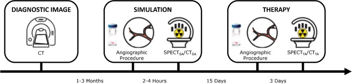
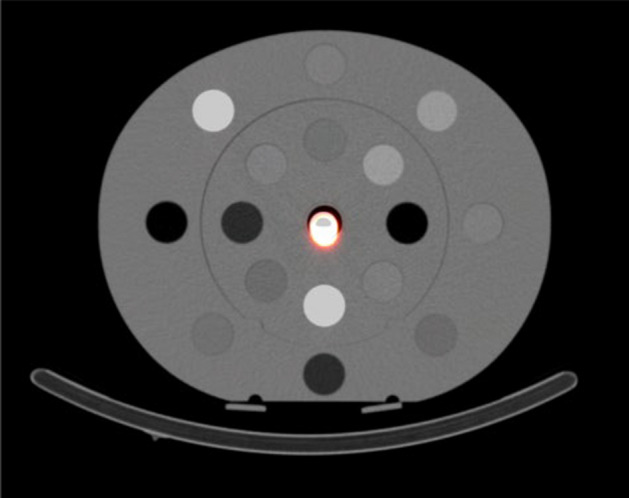
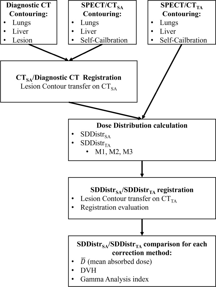
Methods: A 1.9 GBq 166Ho-PLLA activity source was placed in a CIRS phantom and imaged over a week until activity reached 83 MBq, assessing DT effects. Fifteen patients with a single hepatic lesion underwent SA and TA two weeks apart with following SPECT/CT imaging. The mean absorbed dose (AD) and distribution were calculated using the Local Energy Deposition (LED) method for liver, healthy liver (HL) and tumor contours. Three methods were compared for TA AD estimation: no DT correction (M1), whole-image DT correction (M2), and DT correction only for tumor ROI counts (M3). Linear correlation and percentage differences (ΔD%) between SA and TA AD were analyzed. AD distributions in SA and TA were rigidly registered for gamma index analysis (Dose Difference of 10% and Distance to Agreement of 10 mm).
Results: DT effects were significant for activity above 250 MBq (> 11.5%). Strong linear correlations between mean AD values in SA and TA were observed across methods. ΔD% between SA and TA for the liver contour was - 8.6% (M1), 21.5% (M2), and 8.2% (M3). For the HL contour, ΔD% was 8.1% (M1) and 39.0% (M2), while for the tumor contour, it was - 20.1% (M1) and 0.0% (M2). Gamma index pass rates for the liver contour were 76% (M1), 89% (M2), and 92% (M3); for the HL contour, 80% (M1) and 75% (M2); and for the tumor contour, 70% (M1) and 87% (M2).
Conclusion: DT significantly affects TA dose estimation, particularly in tumors. Proper DT correction improves the accuracy of dosimetric evaluation of 166Ho-PLLA for TA in liver and metastases, yielding dose values closer to those obtained in SA, despite the latter not being corrected for DT.
期刊介绍:
EJNMMI Physics is an international platform for scientists, users and adopters of nuclear medicine with a particular interest in physics matters. As a companion journal to the European Journal of Nuclear Medicine and Molecular Imaging, this journal has a multi-disciplinary approach and welcomes original materials and studies with a focus on applied physics and mathematics as well as imaging systems engineering and prototyping in nuclear medicine. This includes physics-driven approaches or algorithms supported by physics that foster early clinical adoption of nuclear medicine imaging and therapy.

 求助内容:
求助内容: 应助结果提醒方式:


