Muhamed M Farhan-Alanie, James Miller, Alastair Stephens, Tsun Yu Kwan, Tarek Boutefnouchet
{"title":"The association between surgeon grade and radiographic implant alignment following oxford unicompartmental knee replacement.","authors":"Muhamed M Farhan-Alanie, James Miller, Alastair Stephens, Tsun Yu Kwan, Tarek Boutefnouchet","doi":"10.1007/s00402-025-05973-y","DOIUrl":null,"url":null,"abstract":"<p><strong>Introduction: </strong>Unicompartmental knee replacement (UKR) is a technically challenging operation. Component alignment can influence implant longevity and knee function post-operatively. This study aimed to investigate implant alignment following UKR performed by consultants compared to trainees.</p><p><strong>Methods: </strong>100 Oxford UKRs performed by trainees and consultants were analysed. Two blinded surgeons independently assessed post-operative knee radiographs on four parameters: flexion/extension of femoral component, posterior slope of tibial component, and varus/valgus of femoral and tibial components. Logistic regression was performed to predict the probability of implant malpositioning outside the optimal position range.</p><p><strong>Results: </strong>Median varus/valgus angles for femoral components did not differ significantly between trainees and consultants (p = 0.92), nor did the angles for tibial components (p = 0.43). Posterior tibial slope measurements showed a significant difference between trainees and consultants (7.08° [IQR 5.2-9.30], and 5.35° [IQR 2.65-7.05], respectively; p < 0.01). Median flexion/extension angles for femoral components also differed significantly between trainees and consultants (-14.45° [IQR -19.2 to -9.85] and -10.2°[IQR -13.55 to -6.95], respectively; p < 0.01). A greater proportion of implants positioned by trainees were classified as outliers for this parameter (46% versus 20%, p < 0.01; aOR 5.39, 95% CI 2.05-14.18, p < 0.01). However, no differences in the proportion of outliers was found when trainees were directly supervised by consultants (p = 0.73).</p><p><strong>Conclusions: </strong>Trainees achieved adequate component alignment within optimal ranges for most parameters however were more prone to positioning the femoral component in excessive flexion. Greater emphasis on achieving optimal flexion/extension positioning of the femoral component during surgical training and direct supervision may improve the outcomes of patients undergoing an Oxford UKR by trainees.</p>","PeriodicalId":8326,"journal":{"name":"Archives of Orthopaedic and Trauma Surgery","volume":"145 1","pages":"362"},"PeriodicalIF":2.1000,"publicationDate":"2025-07-03","publicationTypes":"Journal Article","fieldsOfStudy":null,"isOpenAccess":false,"openAccessPdf":"https://www.ncbi.nlm.nih.gov/pmc/articles/PMC12226701/pdf/","citationCount":"0","resultStr":null,"platform":"Semanticscholar","paperid":null,"PeriodicalName":"Archives of Orthopaedic and Trauma Surgery","FirstCategoryId":"3","ListUrlMain":"https://doi.org/10.1007/s00402-025-05973-y","RegionNum":3,"RegionCategory":"医学","ArticlePicture":[],"TitleCN":null,"AbstractTextCN":null,"PMCID":null,"EPubDate":"","PubModel":"","JCR":"Q2","JCRName":"ORTHOPEDICS","Score":null,"Total":0}
引用次数: 0
Abstract
Introduction: Unicompartmental knee replacement (UKR) is a technically challenging operation. Component alignment can influence implant longevity and knee function post-operatively. This study aimed to investigate implant alignment following UKR performed by consultants compared to trainees.
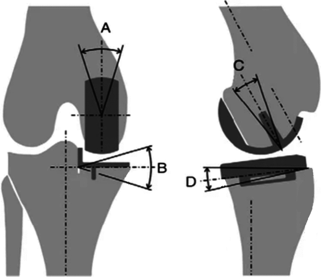
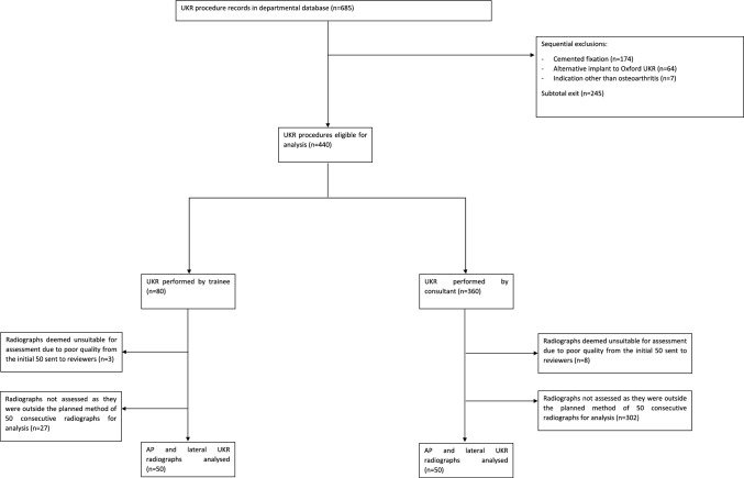
Methods: 100 Oxford UKRs performed by trainees and consultants were analysed. Two blinded surgeons independently assessed post-operative knee radiographs on four parameters: flexion/extension of femoral component, posterior slope of tibial component, and varus/valgus of femoral and tibial components. Logistic regression was performed to predict the probability of implant malpositioning outside the optimal position range.
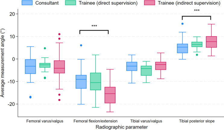
Results: Median varus/valgus angles for femoral components did not differ significantly between trainees and consultants (p = 0.92), nor did the angles for tibial components (p = 0.43). Posterior tibial slope measurements showed a significant difference between trainees and consultants (7.08° [IQR 5.2-9.30], and 5.35° [IQR 2.65-7.05], respectively; p < 0.01). Median flexion/extension angles for femoral components also differed significantly between trainees and consultants (-14.45° [IQR -19.2 to -9.85] and -10.2°[IQR -13.55 to -6.95], respectively; p < 0.01). A greater proportion of implants positioned by trainees were classified as outliers for this parameter (46% versus 20%, p < 0.01; aOR 5.39, 95% CI 2.05-14.18, p < 0.01). However, no differences in the proportion of outliers was found when trainees were directly supervised by consultants (p = 0.73).
Conclusions: Trainees achieved adequate component alignment within optimal ranges for most parameters however were more prone to positioning the femoral component in excessive flexion. Greater emphasis on achieving optimal flexion/extension positioning of the femoral component during surgical training and direct supervision may improve the outcomes of patients undergoing an Oxford UKR by trainees.
期刊介绍:
"Archives of Orthopaedic and Trauma Surgery" is a rich source of instruction and information for physicians in clinical practice and research in the extensive field of orthopaedics and traumatology. The journal publishes papers that deal with diseases and injuries of the musculoskeletal system from all fields and aspects of medicine. The journal is particularly interested in papers that satisfy the information needs of orthopaedic clinicians and practitioners. The journal places special emphasis on clinical relevance.
"Archives of Orthopaedic and Trauma Surgery" is the official journal of the German Speaking Arthroscopy Association (AGA).
 求助内容:
求助内容: 应助结果提醒方式:


