John D Wright, Isaline Renard, Isis A Middleton, Juozas Domarkas, Émer M Foyle, Paul J Lusby, Stephen J Archibald
{"title":"Quantitative whole-body dynamic planar scintigraphy in mice with <sup>99m</sup>Tc and <sup>161</sup>Tb.","authors":"John D Wright, Isaline Renard, Isis A Middleton, Juozas Domarkas, Émer M Foyle, Paul J Lusby, Stephen J Archibald","doi":"10.1186/s40658-025-00775-y","DOIUrl":null,"url":null,"abstract":"<p><strong>Background: </strong>Planar scintigraphy remains commonplace in clinical practice and has been used for quantification and dosimetry estimation over an expanding range of gamma-emitting radionuclides in recent years. Applications of planar scintigraphy, in combination with SPECT/CT imaging, can add value to radiopharmaceutical development in preclinical models and in translation to human use. The aim of this study was to demonstrate whole-body quantitative accuracy in mice using pinhole collimated planar scintigraphy on a preclinical SPECT/CT system, following corrections to sensitivity variations across the field of view.</p><p><strong>Results: </strong>Planar projections were acquired using short imaging time frames, thus allowing for dynamic biodistribution data to be collected and compared to the known injected activity and whole-body SPECT data. Encapsulation of [<sup>99m</sup>Tc]TcO<sub>4</sub><sup>-</sup> in a supramolecular cage was used to demonstrate the visual and quantitative changes in biodistribution over time, as compared to [<sup>99m</sup>Tc]TcO<sub>4</sub><sup>-</sup> alone. For these radiopharmaceuticals, whole-body quantification was 98.7 ± 7.3% of the decay-corrected true injected activity, as opposed to 74.8 ± 7.5% when calculated without a sensitivity correction. Similarly, the final planar scintigraphy frame acquired at 1-hour post-injection quantitatively agreed with activity values returned from the whole-body SPECT: 99.5 ± 10.6% (final frame, planar) vs. 99.1 ± 5.5% (SPECT). Regions of interest (ROIs) over selected organs between planar scintigraphy and SPECT were also in good agreement. Quantitative accuracy of planar scintigraphy was further validated in a preclinical tumour model of prostate cancer using [<sup>161</sup>Tb]Tb-PSMA-617. In this case, the whole-body planar value was 94.6 ± 3.6% of the recorded injected activity and, consistent with <sup>99m</sup>Tc findings, was underestimated without sensitivity correction (76.6 ± 3.1%). Tumour uptake values were equivalent between corrected planar scintigraphy (5.2%IA) and SPECT (5.3%IA) at 1-hour post-injection.</p><p><strong>Conclusions: </strong>Using a common radionuclide and one of emerging radiotherapeutic interest, whole-body injected activity and organ-specific ROI values obtained by planar scintigraphy strongly correlated to the true injected activity and values obtained by SPECT following sensitivity-based corrections. The addition of quantitative dynamic planar scintigraphy into the preclinical workflow followed by SPECT imaging adds value to pharmacokinetic and dosimetry assessments of novel gamma-emitting radiopharmaceuticals in imaging and therapeutic applications.</p>","PeriodicalId":11559,"journal":{"name":"EJNMMI Physics","volume":"12 1","pages":"61"},"PeriodicalIF":3.2000,"publicationDate":"2025-07-01","publicationTypes":"Journal Article","fieldsOfStudy":null,"isOpenAccess":false,"openAccessPdf":"https://www.ncbi.nlm.nih.gov/pmc/articles/PMC12214159/pdf/","citationCount":"0","resultStr":null,"platform":"Semanticscholar","paperid":null,"PeriodicalName":"EJNMMI Physics","FirstCategoryId":"3","ListUrlMain":"https://doi.org/10.1186/s40658-025-00775-y","RegionNum":2,"RegionCategory":"医学","ArticlePicture":[],"TitleCN":null,"AbstractTextCN":null,"PMCID":null,"EPubDate":"","PubModel":"","JCR":"Q2","JCRName":"RADIOLOGY, NUCLEAR MEDICINE & MEDICAL IMAGING","Score":null,"Total":0}
引用次数: 0
Abstract
Background: Planar scintigraphy remains commonplace in clinical practice and has been used for quantification and dosimetry estimation over an expanding range of gamma-emitting radionuclides in recent years. Applications of planar scintigraphy, in combination with SPECT/CT imaging, can add value to radiopharmaceutical development in preclinical models and in translation to human use. The aim of this study was to demonstrate whole-body quantitative accuracy in mice using pinhole collimated planar scintigraphy on a preclinical SPECT/CT system, following corrections to sensitivity variations across the field of view.
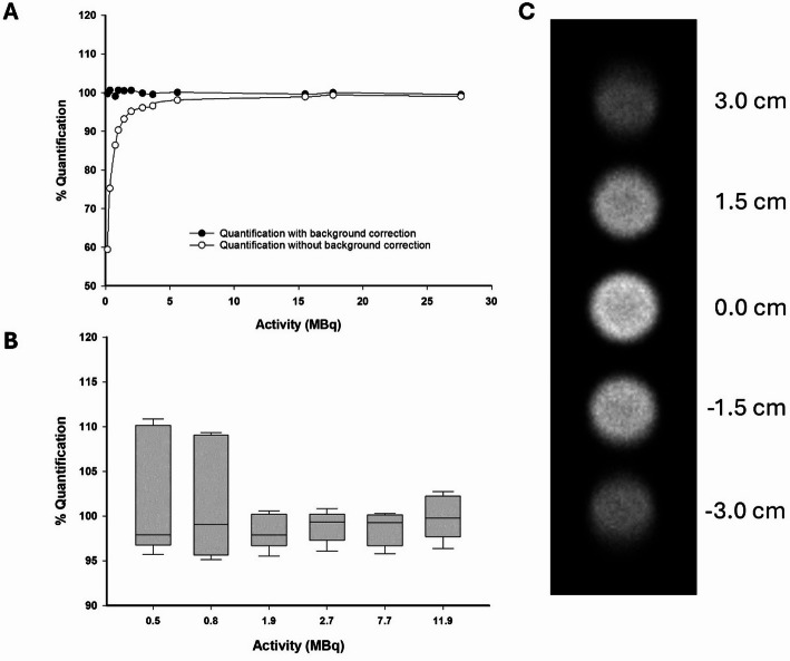
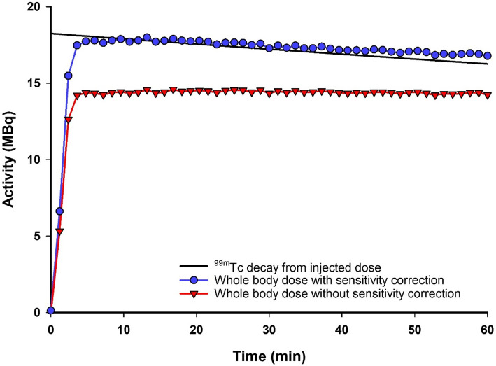
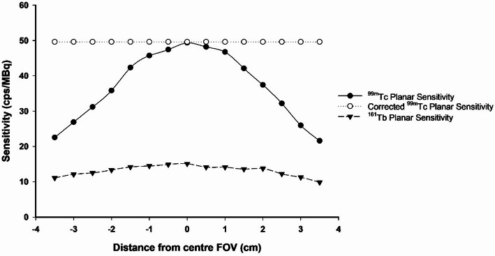
Results: Planar projections were acquired using short imaging time frames, thus allowing for dynamic biodistribution data to be collected and compared to the known injected activity and whole-body SPECT data. Encapsulation of [99mTc]TcO4- in a supramolecular cage was used to demonstrate the visual and quantitative changes in biodistribution over time, as compared to [99mTc]TcO4- alone. For these radiopharmaceuticals, whole-body quantification was 98.7 ± 7.3% of the decay-corrected true injected activity, as opposed to 74.8 ± 7.5% when calculated without a sensitivity correction. Similarly, the final planar scintigraphy frame acquired at 1-hour post-injection quantitatively agreed with activity values returned from the whole-body SPECT: 99.5 ± 10.6% (final frame, planar) vs. 99.1 ± 5.5% (SPECT). Regions of interest (ROIs) over selected organs between planar scintigraphy and SPECT were also in good agreement. Quantitative accuracy of planar scintigraphy was further validated in a preclinical tumour model of prostate cancer using [161Tb]Tb-PSMA-617. In this case, the whole-body planar value was 94.6 ± 3.6% of the recorded injected activity and, consistent with 99mTc findings, was underestimated without sensitivity correction (76.6 ± 3.1%). Tumour uptake values were equivalent between corrected planar scintigraphy (5.2%IA) and SPECT (5.3%IA) at 1-hour post-injection.
Conclusions: Using a common radionuclide and one of emerging radiotherapeutic interest, whole-body injected activity and organ-specific ROI values obtained by planar scintigraphy strongly correlated to the true injected activity and values obtained by SPECT following sensitivity-based corrections. The addition of quantitative dynamic planar scintigraphy into the preclinical workflow followed by SPECT imaging adds value to pharmacokinetic and dosimetry assessments of novel gamma-emitting radiopharmaceuticals in imaging and therapeutic applications.
期刊介绍:
EJNMMI Physics is an international platform for scientists, users and adopters of nuclear medicine with a particular interest in physics matters. As a companion journal to the European Journal of Nuclear Medicine and Molecular Imaging, this journal has a multi-disciplinary approach and welcomes original materials and studies with a focus on applied physics and mathematics as well as imaging systems engineering and prototyping in nuclear medicine. This includes physics-driven approaches or algorithms supported by physics that foster early clinical adoption of nuclear medicine imaging and therapy.

 求助内容:
求助内容: 应助结果提醒方式:


