Alistair Weld, Luke Dixon, Michael Dyck, Giulio Anichini, Alex Ranne, Sophie Camp, Stamatia Giannarou
{"title":"Identifying visible tissue in intraoperative ultrasound: a method and application.","authors":"Alistair Weld, Luke Dixon, Michael Dyck, Giulio Anichini, Alex Ranne, Sophie Camp, Stamatia Giannarou","doi":"10.1007/s11548-025-03415-z","DOIUrl":null,"url":null,"abstract":"<p><strong>Purpose: </strong>Intraoperative ultrasound scanning is a demanding visuotactile task. It requires operators to simultaneously localise the ultrasound perspective and manually perform slight adjustments to the pose of the probe, making sure not to apply excessive force or breaking contact with the tissue, while also characterising the visible tissue.</p><p><strong>Method: </strong>To analyse the probe-tissue contact, an iterative filtering and topological method is proposed to identify the underlying visible tissue, which can be used to detect acoustic shadow and construct confidence maps of perceptual salience.</p><p><strong>Results: </strong>For evaluation, datasets containing both in vivo and medical phantom data are created. A suite of evaluations is performed, including an evaluation of acoustic shadow classification. Compared to an ablation, deep learning, and statistical method, the proposed approach achieves superior classification on in vivo data, achieving an <math><msub><mi>F</mi> <mi>β</mi></msub> </math> score of 0.864, in comparison with 0.838, 0.808, and 0.808. A novel framework for evaluating the confidence estimation of probe-tissue contact is created. The phantom data are captured specifically for this, and comparison is made against two established methods. The proposed method produced the superior response, achieving an average normalised root-mean-square error of 0.168, in comparison with 1.836 and 4.542. Evaluation is also extended to determine the algorithm's robustness to parameter perturbation, speckle noise, data distribution shift, and capability for guiding a robotic scan.</p><p><strong>Conclusion: </strong>The results of this comprehensive set of experiments justify the potential clinical value of the proposed algorithm, which can be used to support clinical training and robotic ultrasound automation.</p>","PeriodicalId":51251,"journal":{"name":"International Journal of Computer Assisted Radiology and Surgery","volume":" ","pages":"2107-2117"},"PeriodicalIF":2.3000,"publicationDate":"2025-10-01","publicationTypes":"Journal Article","fieldsOfStudy":null,"isOpenAccess":false,"openAccessPdf":"https://www.ncbi.nlm.nih.gov/pmc/articles/PMC12518381/pdf/","citationCount":"0","resultStr":null,"platform":"Semanticscholar","paperid":null,"PeriodicalName":"International Journal of Computer Assisted Radiology and Surgery","FirstCategoryId":"5","ListUrlMain":"https://doi.org/10.1007/s11548-025-03415-z","RegionNum":3,"RegionCategory":"医学","ArticlePicture":[],"TitleCN":null,"AbstractTextCN":null,"PMCID":null,"EPubDate":"2025/6/28 0:00:00","PubModel":"Epub","JCR":"Q3","JCRName":"ENGINEERING, BIOMEDICAL","Score":null,"Total":0}
引用次数: 0
Abstract
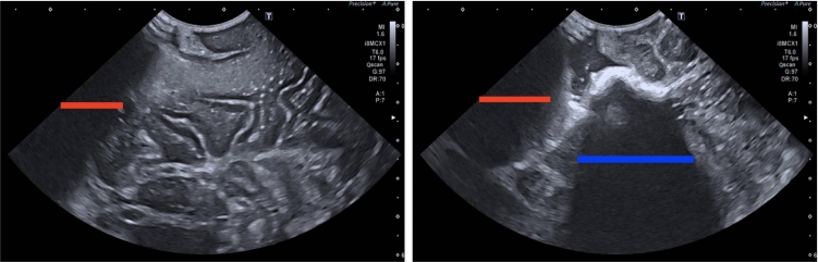
Purpose: Intraoperative ultrasound scanning is a demanding visuotactile task. It requires operators to simultaneously localise the ultrasound perspective and manually perform slight adjustments to the pose of the probe, making sure not to apply excessive force or breaking contact with the tissue, while also characterising the visible tissue.
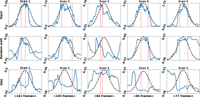
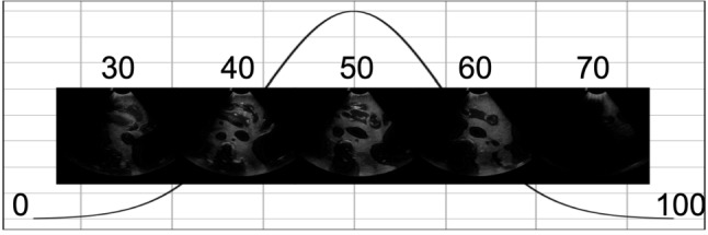
Method: To analyse the probe-tissue contact, an iterative filtering and topological method is proposed to identify the underlying visible tissue, which can be used to detect acoustic shadow and construct confidence maps of perceptual salience.
Results: For evaluation, datasets containing both in vivo and medical phantom data are created. A suite of evaluations is performed, including an evaluation of acoustic shadow classification. Compared to an ablation, deep learning, and statistical method, the proposed approach achieves superior classification on in vivo data, achieving an score of 0.864, in comparison with 0.838, 0.808, and 0.808. A novel framework for evaluating the confidence estimation of probe-tissue contact is created. The phantom data are captured specifically for this, and comparison is made against two established methods. The proposed method produced the superior response, achieving an average normalised root-mean-square error of 0.168, in comparison with 1.836 and 4.542. Evaluation is also extended to determine the algorithm's robustness to parameter perturbation, speckle noise, data distribution shift, and capability for guiding a robotic scan.
Conclusion: The results of this comprehensive set of experiments justify the potential clinical value of the proposed algorithm, which can be used to support clinical training and robotic ultrasound automation.
期刊介绍:
The International Journal for Computer Assisted Radiology and Surgery (IJCARS) is a peer-reviewed journal that provides a platform for closing the gap between medical and technical disciplines, and encourages interdisciplinary research and development activities in an international environment.

 求助内容:
求助内容: 应助结果提醒方式:


