Sharara Fadhil Abbood, Israa Jawad Abdul-Rasul, Amaal Sahib Al-Zughaibi, Hamzah H Kzar Al-Shukri, Fadak B Rabee, Zainab A Fadhil, Zainab A A Radhi
{"title":"Comparison of Brain Activity between Patients with Parkinson Disease Dementia and Patients Affected by Dementia with Lewy Body through EEG Analysis.","authors":"Sharara Fadhil Abbood, Israa Jawad Abdul-Rasul, Amaal Sahib Al-Zughaibi, Hamzah H Kzar Al-Shukri, Fadak B Rabee, Zainab A Fadhil, Zainab A A Radhi","doi":"10.18502/ijps.v19i4.16548","DOIUrl":null,"url":null,"abstract":"<p><p><b>Objective:</b> Parkinson's disease dementia (PDD) and Dementia with Lewy bodies (DLB) are two syndromes categorized under synucleinopathy, sharing comparable symptoms. The identification of biomarkers would offer an accurate approach for improved diagnosis, treatment, and monitoring of treatment efficacy for these distinct forms of dementia. <b>Method</b> <b>:</b> This study utilized spectral analysis and nonlinear dynamic analysis to compare electroencephalogram (EEG) characteristics between PDD and DLB patients. EEG data was collected from 30 PDD patients, 36 DLB patients, and 36 healthy subjects at rest. Following a conditioning phase to minimize noise and eliminate artifacts, we derived spectral and complexity features using Welch's method and sample entropy. Analysis of variance with repeated measures was performed to compare spectral features and nonlinear dynamics of brain activity between the groups. <b>Results:</b> Post hoc comparison showed that in the control group, the power of delta and theta bands was lower and the power of alpha and beta bands was higher than in patients with PDD and DLB. (P < 0.05). In the theta and alpha bands, the PDD group showed greater power than the DLB group (P < 0.05). Furthermore, there was a significant main effect of diagnosis (F = 4.67, P = 0.007), and also the diagnosis by region interaction for complexity values (F = 4.58, P = 0.009). Post hoc analysis showed that the EEG complexity of the control group was significantly higher than that of the PDD and DLB groups in the frontal, central, temporal and parietal regions (P < 0.05). Moreover, the EEG complexity of the PDD group was significantly higher than that of the DLB group in the central, temporal and parietal regions (P < 0.05). <b>Conclusion:</b> Although both PDD and DLB had almost similar patterns compared to the control group, they showed differences in the EEG power spectrum and its nonlinear dynamics. Our findings indicated marked diffuse slowing and lower cortical complexity or activity in DLB patients compared to PDD in all regions, especially in the central, temporal and parietal areas.</p>","PeriodicalId":38866,"journal":{"name":"Iranian Journal of Psychiatry","volume":"19 4","pages":"346-355"},"PeriodicalIF":0.0000,"publicationDate":"2024-10-01","publicationTypes":"Journal Article","fieldsOfStudy":null,"isOpenAccess":false,"openAccessPdf":"https://www.ncbi.nlm.nih.gov/pmc/articles/PMC12206469/pdf/","citationCount":"0","resultStr":null,"platform":"Semanticscholar","paperid":null,"PeriodicalName":"Iranian Journal of Psychiatry","FirstCategoryId":"1085","ListUrlMain":"https://doi.org/10.18502/ijps.v19i4.16548","RegionNum":0,"RegionCategory":null,"ArticlePicture":[],"TitleCN":null,"AbstractTextCN":null,"PMCID":null,"EPubDate":"","PubModel":"","JCR":"Q2","JCRName":"Medicine","Score":null,"Total":0}
引用次数: 0
Abstract
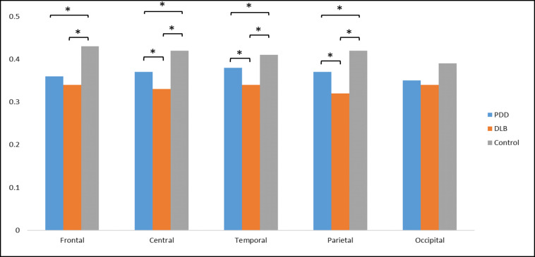
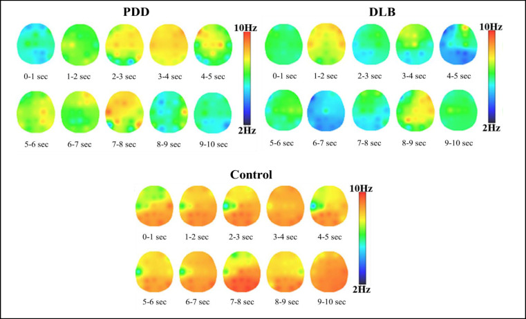
Objective: Parkinson's disease dementia (PDD) and Dementia with Lewy bodies (DLB) are two syndromes categorized under synucleinopathy, sharing comparable symptoms. The identification of biomarkers would offer an accurate approach for improved diagnosis, treatment, and monitoring of treatment efficacy for these distinct forms of dementia. Method: This study utilized spectral analysis and nonlinear dynamic analysis to compare electroencephalogram (EEG) characteristics between PDD and DLB patients. EEG data was collected from 30 PDD patients, 36 DLB patients, and 36 healthy subjects at rest. Following a conditioning phase to minimize noise and eliminate artifacts, we derived spectral and complexity features using Welch's method and sample entropy. Analysis of variance with repeated measures was performed to compare spectral features and nonlinear dynamics of brain activity between the groups. Results: Post hoc comparison showed that in the control group, the power of delta and theta bands was lower and the power of alpha and beta bands was higher than in patients with PDD and DLB. (P < 0.05). In the theta and alpha bands, the PDD group showed greater power than the DLB group (P < 0.05). Furthermore, there was a significant main effect of diagnosis (F = 4.67, P = 0.007), and also the diagnosis by region interaction for complexity values (F = 4.58, P = 0.009). Post hoc analysis showed that the EEG complexity of the control group was significantly higher than that of the PDD and DLB groups in the frontal, central, temporal and parietal regions (P < 0.05). Moreover, the EEG complexity of the PDD group was significantly higher than that of the DLB group in the central, temporal and parietal regions (P < 0.05). Conclusion: Although both PDD and DLB had almost similar patterns compared to the control group, they showed differences in the EEG power spectrum and its nonlinear dynamics. Our findings indicated marked diffuse slowing and lower cortical complexity or activity in DLB patients compared to PDD in all regions, especially in the central, temporal and parietal areas.

 求助内容:
求助内容: 应助结果提醒方式:


