{"title":"Cervical vertebrae fusion in elbow knee synostosis (Eks)-mutant mice with fibroblast growth factor 9 N143T mutation","authors":"Georgina Djameh, Masayo Harada, Keiichi Akita","doi":"10.1111/cga.70016","DOIUrl":null,"url":null,"abstract":"<p>Proper vertebral column development requires precise segmentation and regulated chondrogenesis during embryogenesis. Mutations affecting fibroblast growth factor 9 (FGF9) signaling disrupt these processes, resulting in abnormal vertebral column development. A missense mutation in FGF9 (p.Asn143Thr) produces <i>elbow knee synostosis</i> (<i>Eks</i>)-mutant mice, which display skeletal fusions, including those in the vertebral column, underscoring the essential role of FGF9 in vertebral segmentation and vertebral joint development. However, the mechanisms regulating joint formation in vertebrae remain elusive. Here, we report that the homozygous <i>Eks</i> mutant mice exhibit neural arch lamina fusion along the rostrocaudal axis at the dorsolateral position in neonates. We investigated the cellular and molecular mechanisms underlying the cervical vertebral fusion in <i>Fgf9</i><sup><i>Eks/Eks</i></sup> embryos. <i>Fgf9</i><sup><i>Eks/Eks</i></sup> embryos showed multiple fusions and thickened cartilage of cervical lamina on embryonic day (E) 14.5 and E13.5. Additionally, <i>Fgf9</i><sup><i>Eks/Eks</i></sup> embryos exhibited COL2A1 expression domain expansion accompanied by ectopic chondrocyte accumulation in the presumptive interlaminar space on E12.5 and E11.5. These anomalies persisted through endochondral ossification, leading to postnatal cervical vertebral bone fusion. Ectopic expression of COL2A1, Cyclin D1, and fibroblast growth factor (FGF) signaling target ETV4 was observed in the presumptive interlaminar space, indicating altered cell proliferation and cell fate specification. These findings demonstrate that FGF9<sup><i>Eks</i></sup> protein interferes with vertebral column segmentation by impairing chondrogenic boundary regulation through ectopic cell proliferation and transcriptional activity. In conclusion, ectopic FGF9 signaling leads to cervical vertebral fusion, highlighting its contributing role in maintaining vertebral segmentation and chondrogenesis during embryogenesis.</p>","PeriodicalId":10626,"journal":{"name":"Congenital Anomalies","volume":"65 1","pages":""},"PeriodicalIF":1.6000,"publicationDate":"2025-07-01","publicationTypes":"Journal Article","fieldsOfStudy":null,"isOpenAccess":false,"openAccessPdf":"https://onlinelibrary.wiley.com/doi/epdf/10.1111/cga.70016","citationCount":"0","resultStr":null,"platform":"Semanticscholar","paperid":null,"PeriodicalName":"Congenital Anomalies","FirstCategoryId":"3","ListUrlMain":"https://onlinelibrary.wiley.com/doi/10.1111/cga.70016","RegionNum":4,"RegionCategory":"医学","ArticlePicture":[],"TitleCN":null,"AbstractTextCN":null,"PMCID":null,"EPubDate":"","PubModel":"","JCR":"Q3","JCRName":"PEDIATRICS","Score":null,"Total":0}
引用次数: 0
Abstract
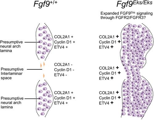
Proper vertebral column development requires precise segmentation and regulated chondrogenesis during embryogenesis. Mutations affecting fibroblast growth factor 9 (FGF9) signaling disrupt these processes, resulting in abnormal vertebral column development. A missense mutation in FGF9 (p.Asn143Thr) produces elbow knee synostosis (Eks)-mutant mice, which display skeletal fusions, including those in the vertebral column, underscoring the essential role of FGF9 in vertebral segmentation and vertebral joint development. However, the mechanisms regulating joint formation in vertebrae remain elusive. Here, we report that the homozygous Eks mutant mice exhibit neural arch lamina fusion along the rostrocaudal axis at the dorsolateral position in neonates. We investigated the cellular and molecular mechanisms underlying the cervical vertebral fusion in Fgf9Eks/Eks embryos. Fgf9Eks/Eks embryos showed multiple fusions and thickened cartilage of cervical lamina on embryonic day (E) 14.5 and E13.5. Additionally, Fgf9Eks/Eks embryos exhibited COL2A1 expression domain expansion accompanied by ectopic chondrocyte accumulation in the presumptive interlaminar space on E12.5 and E11.5. These anomalies persisted through endochondral ossification, leading to postnatal cervical vertebral bone fusion. Ectopic expression of COL2A1, Cyclin D1, and fibroblast growth factor (FGF) signaling target ETV4 was observed in the presumptive interlaminar space, indicating altered cell proliferation and cell fate specification. These findings demonstrate that FGF9Eks protein interferes with vertebral column segmentation by impairing chondrogenic boundary regulation through ectopic cell proliferation and transcriptional activity. In conclusion, ectopic FGF9 signaling leads to cervical vertebral fusion, highlighting its contributing role in maintaining vertebral segmentation and chondrogenesis during embryogenesis.
期刊介绍:
Congenital Anomalies is the official English language journal of the Japanese Teratology Society, and publishes original articles in laboratory as well as clinical research in all areas of abnormal development and related fields, from all over the world. Although contributions by members of the teratology societies affiliated with The International Federation of Teratology Societies are given priority, contributions from non-members are welcomed.
 求助内容:
求助内容: 应助结果提醒方式:


