Qianhe Liu, Jiahui Jiang, Kewei Wu, Yan Zhang, Nan Sun, Jiawen Luo, Te Ba, Aiqing Lv, Chuane Liu, Yiyu Yin, Zhenghan Yang, Hui Xu
{"title":"A two-step automatic identification of contrast phases for abdominal CT images based on residual networks.","authors":"Qianhe Liu, Jiahui Jiang, Kewei Wu, Yan Zhang, Nan Sun, Jiawen Luo, Te Ba, Aiqing Lv, Chuane Liu, Yiyu Yin, Zhenghan Yang, Hui Xu","doi":"10.1186/s13244-025-01995-7","DOIUrl":null,"url":null,"abstract":"<p><strong>Objectives: </strong>To develop a deep learning model based on Residual Networks (ResNet) for the automated and accurate identification of contrast phases in abdominal CT images.</p><p><strong>Methods: </strong>A dataset of 1175 abdominal contrast-enhanced CT scans was retrospectively collected for the model development, and another independent dataset of 215 scans from five hospitals was collected for external testing. Each contrast phase was independently annotated by two radiologists. A ResNet-based model was developed to automatically classify phases into the early arterial phase (EAP) or late arterial phase (LAP), portal venous phase (PVP), and delayed phase (DP). Strategy A identified EAP or LAP, PVP, and DP in one step. Strategy B used a two-step approach: first classifying images as arterial phase (AP), PVP, and DP, then further classifying AP images into EAP or LAP. Model performance and strategy comparison were evaluated.</p><p><strong>Results: </strong>In the internal test set, the overall accuracy of the two-step strategy was 98.3% (283/288; p < 0.001), significantly higher than that of the one-step strategy (91.7%, 264/288; p < 0.001). In the external test set, the two-step model achieved an overall accuracy of 99.1% (639/645), with sensitivities of 95.1% (EAP), 99.4% (LAP), 99.5% (PVP), and 99.5% (DP).</p><p><strong>Conclusion: </strong>The proposed two-step ResNet-based model provides highly accurate and robust identification of contrast phases in abdominal CT images, outperforming the conventional one-step strategy.</p><p><strong>Critical relevance statement: </strong>Automated and accurate identification of contrast phases in abdominal CT images provides a robust tool for improving image quality control and establishes a strong foundation for AI-driven applications, particularly those leveraging contrast-enhanced abdominal imaging data.</p><p><strong>Key points: </strong>Accurate identification of contrast phases is crucial in abdominal CT imaging. The two-step ResNet-based model achieved superior accuracy across internal and external datasets. Automated phase classification strengthens imaging quality control and supports precision AI applications.</p>","PeriodicalId":13639,"journal":{"name":"Insights into Imaging","volume":"16 1","pages":"139"},"PeriodicalIF":4.5000,"publicationDate":"2025-06-27","publicationTypes":"Journal Article","fieldsOfStudy":null,"isOpenAccess":false,"openAccessPdf":"https://www.ncbi.nlm.nih.gov/pmc/articles/PMC12204963/pdf/","citationCount":"0","resultStr":null,"platform":"Semanticscholar","paperid":null,"PeriodicalName":"Insights into Imaging","FirstCategoryId":"3","ListUrlMain":"https://doi.org/10.1186/s13244-025-01995-7","RegionNum":2,"RegionCategory":"医学","ArticlePicture":[],"TitleCN":null,"AbstractTextCN":null,"PMCID":null,"EPubDate":"","PubModel":"","JCR":"Q1","JCRName":"RADIOLOGY, NUCLEAR MEDICINE & MEDICAL IMAGING","Score":null,"Total":0}
引用次数: 0
Abstract
Objectives: To develop a deep learning model based on Residual Networks (ResNet) for the automated and accurate identification of contrast phases in abdominal CT images.
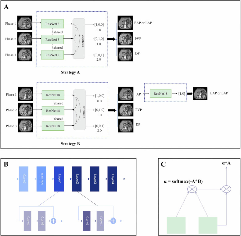
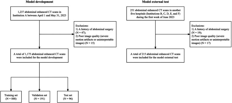
Methods: A dataset of 1175 abdominal contrast-enhanced CT scans was retrospectively collected for the model development, and another independent dataset of 215 scans from five hospitals was collected for external testing. Each contrast phase was independently annotated by two radiologists. A ResNet-based model was developed to automatically classify phases into the early arterial phase (EAP) or late arterial phase (LAP), portal venous phase (PVP), and delayed phase (DP). Strategy A identified EAP or LAP, PVP, and DP in one step. Strategy B used a two-step approach: first classifying images as arterial phase (AP), PVP, and DP, then further classifying AP images into EAP or LAP. Model performance and strategy comparison were evaluated.
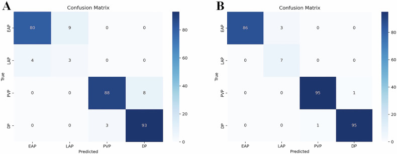
Results: In the internal test set, the overall accuracy of the two-step strategy was 98.3% (283/288; p < 0.001), significantly higher than that of the one-step strategy (91.7%, 264/288; p < 0.001). In the external test set, the two-step model achieved an overall accuracy of 99.1% (639/645), with sensitivities of 95.1% (EAP), 99.4% (LAP), 99.5% (PVP), and 99.5% (DP).
Conclusion: The proposed two-step ResNet-based model provides highly accurate and robust identification of contrast phases in abdominal CT images, outperforming the conventional one-step strategy.
Critical relevance statement: Automated and accurate identification of contrast phases in abdominal CT images provides a robust tool for improving image quality control and establishes a strong foundation for AI-driven applications, particularly those leveraging contrast-enhanced abdominal imaging data.
Key points: Accurate identification of contrast phases is crucial in abdominal CT imaging. The two-step ResNet-based model achieved superior accuracy across internal and external datasets. Automated phase classification strengthens imaging quality control and supports precision AI applications.
期刊介绍:
Insights into Imaging (I³) is a peer-reviewed open access journal published under the brand SpringerOpen. All content published in the journal is freely available online to anyone, anywhere!
I³ continuously updates scientific knowledge and progress in best-practice standards in radiology through the publication of original articles and state-of-the-art reviews and opinions, along with recommendations and statements from the leading radiological societies in Europe.
Founded by the European Society of Radiology (ESR), I³ creates a platform for educational material, guidelines and recommendations, and a forum for topics of controversy.
A balanced combination of review articles, original papers, short communications from European radiological congresses and information on society matters makes I³ an indispensable source for current information in this field.
I³ is owned by the ESR, however authors retain copyright to their article according to the Creative Commons Attribution License (see Copyright and License Agreement). All articles can be read, redistributed and reused for free, as long as the author of the original work is cited properly.
The open access fees (article-processing charges) for this journal are kindly sponsored by ESR for all Members.
The journal went open access in 2012, which means that all articles published since then are freely available online.

 求助内容:
求助内容: 应助结果提醒方式:


