Liwen Zhu, Ben Zhao, Tianyi Xia, Di Chang, Cong Xia, Mengqiu Liu, Ridong Li, Buyue Cao, Yue Qiu, Yaoyao Yu, Shuwei Zhou, Huayu Chen, Wu Cai, Zhimin Ding, Chunqiang Lu, Tianyu Tang, Yang Song, Yuancheng Wang, Jing Ye, Ying Liu, Shenghong Ju
{"title":"A radiomics-based model for predicting lymph nodes metastasis of pancreatic ductal adenocarcinoma: a multicenter study.","authors":"Liwen Zhu, Ben Zhao, Tianyi Xia, Di Chang, Cong Xia, Mengqiu Liu, Ridong Li, Buyue Cao, Yue Qiu, Yaoyao Yu, Shuwei Zhou, Huayu Chen, Wu Cai, Zhimin Ding, Chunqiang Lu, Tianyu Tang, Yang Song, Yuancheng Wang, Jing Ye, Ying Liu, Shenghong Ju","doi":"10.1186/s13244-025-02025-2","DOIUrl":null,"url":null,"abstract":"<p><strong>Purpose: </strong>To develop a radiomics model to predict lymph nodes metastasis (LNM) in patients with pancreatic ductal adenocarcinoma (PDAC) and assess its value for clinical management.</p><p><strong>Methods: </strong>Patients with pathologically confirmed PDAC from four centers were retrospectively enrolled and split into four cohorts: training (n = 192), validation (n = 82), testing (n = 100), and clinical utilization (n = 163). A radiomics model was constructed based on contrast-enhanced CT (CECT) to predict LNM, and its performance was evaluated using the areas under the curve (AUC). Kaplan-Meier analysis was used to assess the prognostic and therapeutic decision-assisting value of the radiomics model.</p><p><strong>Results: </strong>A total of 437 patients (mean age: 63.1 years ± 9.2 standard deviation; 253 men) were included. The radiomics model outperformed other models with AUCs of 0.84, 0.82, and 0.78 in the training, validation, and testing cohorts (all p < 0.05), respectively. LNM predicted by the radiomics model was significantly associated with overall survival (p < 0.001). Kaplan-Meier analysis revealed that patients with a higher risk of LNM also had worse outcomes (all p < 0.05). Additionally, among the high-risk subgroup identified by the radiomics model in the clinical utilization cohort, patients who underwent dissection of ≥ 15 lymph nodes exhibited better overall survival compared to those with fewer lymph nodes dissected (p = 0.002).</p><p><strong>Conclusion: </strong>The radiomics model we constructed demonstrated impressive performance in predicting LNM and prognosis, suggesting its potential for optimizing the clinical management of PDAC.</p><p><strong>Critical relevance statement: </strong>This radiomics model can predict the risk of lymph nodes metastasis and prognosis of patients in pancreatic ductal adenocarcinoma and has potential value in selecting patients who can benefit from different extents of lymph nodes dissection.</p><p><strong>Key points: </strong>Thorough lymph node dissection is important for achieving the best prognosis in pancreatic ductal adenocarcinoma (PDAC). The radiomics model can accurately predict lymph node status and stratify patients' prognosis. This radiomics model enhances the clinical management of PDAC.</p>","PeriodicalId":13639,"journal":{"name":"Insights into Imaging","volume":"16 1","pages":"141"},"PeriodicalIF":4.5000,"publicationDate":"2025-06-27","publicationTypes":"Journal Article","fieldsOfStudy":null,"isOpenAccess":false,"openAccessPdf":"https://www.ncbi.nlm.nih.gov/pmc/articles/PMC12204970/pdf/","citationCount":"0","resultStr":null,"platform":"Semanticscholar","paperid":null,"PeriodicalName":"Insights into Imaging","FirstCategoryId":"3","ListUrlMain":"https://doi.org/10.1186/s13244-025-02025-2","RegionNum":2,"RegionCategory":"医学","ArticlePicture":[],"TitleCN":null,"AbstractTextCN":null,"PMCID":null,"EPubDate":"","PubModel":"","JCR":"Q1","JCRName":"RADIOLOGY, NUCLEAR MEDICINE & MEDICAL IMAGING","Score":null,"Total":0}
引用次数: 0
Abstract
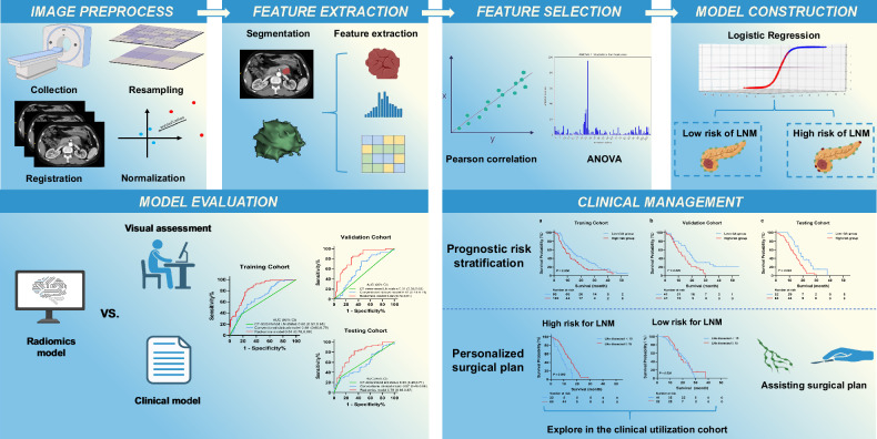
Purpose: To develop a radiomics model to predict lymph nodes metastasis (LNM) in patients with pancreatic ductal adenocarcinoma (PDAC) and assess its value for clinical management.
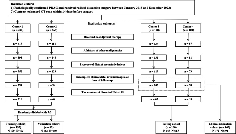
Methods: Patients with pathologically confirmed PDAC from four centers were retrospectively enrolled and split into four cohorts: training (n = 192), validation (n = 82), testing (n = 100), and clinical utilization (n = 163). A radiomics model was constructed based on contrast-enhanced CT (CECT) to predict LNM, and its performance was evaluated using the areas under the curve (AUC). Kaplan-Meier analysis was used to assess the prognostic and therapeutic decision-assisting value of the radiomics model.
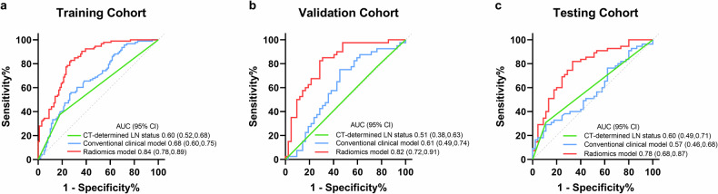
Results: A total of 437 patients (mean age: 63.1 years ± 9.2 standard deviation; 253 men) were included. The radiomics model outperformed other models with AUCs of 0.84, 0.82, and 0.78 in the training, validation, and testing cohorts (all p < 0.05), respectively. LNM predicted by the radiomics model was significantly associated with overall survival (p < 0.001). Kaplan-Meier analysis revealed that patients with a higher risk of LNM also had worse outcomes (all p < 0.05). Additionally, among the high-risk subgroup identified by the radiomics model in the clinical utilization cohort, patients who underwent dissection of ≥ 15 lymph nodes exhibited better overall survival compared to those with fewer lymph nodes dissected (p = 0.002).
Conclusion: The radiomics model we constructed demonstrated impressive performance in predicting LNM and prognosis, suggesting its potential for optimizing the clinical management of PDAC.
Critical relevance statement: This radiomics model can predict the risk of lymph nodes metastasis and prognosis of patients in pancreatic ductal adenocarcinoma and has potential value in selecting patients who can benefit from different extents of lymph nodes dissection.
Key points: Thorough lymph node dissection is important for achieving the best prognosis in pancreatic ductal adenocarcinoma (PDAC). The radiomics model can accurately predict lymph node status and stratify patients' prognosis. This radiomics model enhances the clinical management of PDAC.
期刊介绍:
Insights into Imaging (I³) is a peer-reviewed open access journal published under the brand SpringerOpen. All content published in the journal is freely available online to anyone, anywhere!
I³ continuously updates scientific knowledge and progress in best-practice standards in radiology through the publication of original articles and state-of-the-art reviews and opinions, along with recommendations and statements from the leading radiological societies in Europe.
Founded by the European Society of Radiology (ESR), I³ creates a platform for educational material, guidelines and recommendations, and a forum for topics of controversy.
A balanced combination of review articles, original papers, short communications from European radiological congresses and information on society matters makes I³ an indispensable source for current information in this field.
I³ is owned by the ESR, however authors retain copyright to their article according to the Creative Commons Attribution License (see Copyright and License Agreement). All articles can be read, redistributed and reused for free, as long as the author of the original work is cited properly.
The open access fees (article-processing charges) for this journal are kindly sponsored by ESR for all Members.
The journal went open access in 2012, which means that all articles published since then are freely available online.

 求助内容:
求助内容: 应助结果提醒方式:


