Jingxuan Wang, Xiaowen Zhang, Wei Tang, Marcel van Tuinen, Rozemarijn Vliegenthart, Peter van Ooijen
{"title":"A multi-view CNN model to predict resolving of new lung nodules on follow-up low-dose chest CT.","authors":"Jingxuan Wang, Xiaowen Zhang, Wei Tang, Marcel van Tuinen, Rozemarijn Vliegenthart, Peter van Ooijen","doi":"10.1186/s13244-025-02000-x","DOIUrl":null,"url":null,"abstract":"<p><strong>Objective: </strong>New, intermediate-sized nodules in lung cancer screening undergo follow-up CT, but some of these will resolve. We evaluated the performance of a multi-view convolutional neural network (CNN) in distinguishing resolving and non-resolving new, intermediate-sized lung nodules.</p><p><strong>Materials and methods: </strong>This retrospective study utilized data on 344 intermediate-sized nodules (50-500 mm<sup>3</sup>) in 250 participants from the NELSON (Dutch-Belgian Randomized Lung Cancer Screening) trial. We implemented four-fold cross-validation for model training and testing. A multi-view CNN model was developed by combining three two-dimensional (2D) CNN models and one three-dimensional (3D) CNN model. We used 2D, 2.5D, and 3D models for comparison. The models' performance was evaluated using sensitivity, specificity, and area under the ROC curve (AUC). Specificity, indicating what percentage of non-resolving nodules requiring follow-up can be correctly predicted, was maximized.</p><p><strong>Results: </strong>Among all nodules, 18.3% (63) were resolving. The multi-view CNN model achieved an AUC of 0.81, with a mean sensitivity of 0.63 (SD, 0.15) and a mean specificity of 0.93 (SD, 0.02). The model significantly improved performance compared to 2D, 2.5D, or 3D models (p < 0.05). Under the premise of specificity greater than 90% (meaning < 10% of non-resolving nodules are incorrectly identified as resolving), follow-up CT in 14% of individuals could be prevented.</p><p><strong>Conclusion: </strong>The multi-view CNN model achieved high specificity in discriminating new intermediate nodules that would need follow-up CT by identifying non-resolving nodules. After further validation and optimization, this model may assist with decision-making when new intermediate nodules are found in lung cancer screening.</p><p><strong>Critical relevance statement: </strong>The multi-view CNN-based model has the potential to reduce unnecessary follow-up scans when new nodules are detected, aiding radiologists in making earlier, more informed decisions.</p><p><strong>Key points: </strong>Predicting the resolution of new intermediate lung nodules in lung cancer screening CT is a challenge. Our multi-view CNN model showed an AUC of 0.81, a specificity of 0.93, and a sensitivity of 0.63 at the nodule level. The multi-view model demonstrated a significant improvement in AUC compared to the three 2D models, one 2.5D model, and one 3D model.</p>","PeriodicalId":13639,"journal":{"name":"Insights into Imaging","volume":"16 1","pages":"138"},"PeriodicalIF":4.5000,"publicationDate":"2025-06-27","publicationTypes":"Journal Article","fieldsOfStudy":null,"isOpenAccess":false,"openAccessPdf":"https://www.ncbi.nlm.nih.gov/pmc/articles/PMC12205119/pdf/","citationCount":"0","resultStr":null,"platform":"Semanticscholar","paperid":null,"PeriodicalName":"Insights into Imaging","FirstCategoryId":"3","ListUrlMain":"https://doi.org/10.1186/s13244-025-02000-x","RegionNum":2,"RegionCategory":"医学","ArticlePicture":[],"TitleCN":null,"AbstractTextCN":null,"PMCID":null,"EPubDate":"","PubModel":"","JCR":"Q1","JCRName":"RADIOLOGY, NUCLEAR MEDICINE & MEDICAL IMAGING","Score":null,"Total":0}
引用次数: 0
Abstract
Objective: New, intermediate-sized nodules in lung cancer screening undergo follow-up CT, but some of these will resolve. We evaluated the performance of a multi-view convolutional neural network (CNN) in distinguishing resolving and non-resolving new, intermediate-sized lung nodules.
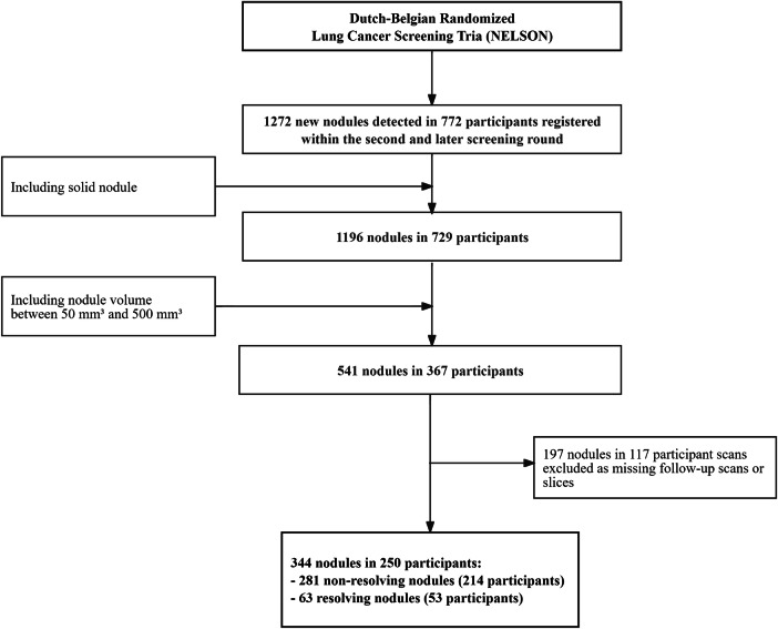
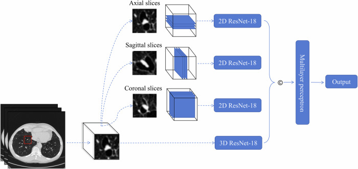
Materials and methods: This retrospective study utilized data on 344 intermediate-sized nodules (50-500 mm3) in 250 participants from the NELSON (Dutch-Belgian Randomized Lung Cancer Screening) trial. We implemented four-fold cross-validation for model training and testing. A multi-view CNN model was developed by combining three two-dimensional (2D) CNN models and one three-dimensional (3D) CNN model. We used 2D, 2.5D, and 3D models for comparison. The models' performance was evaluated using sensitivity, specificity, and area under the ROC curve (AUC). Specificity, indicating what percentage of non-resolving nodules requiring follow-up can be correctly predicted, was maximized.
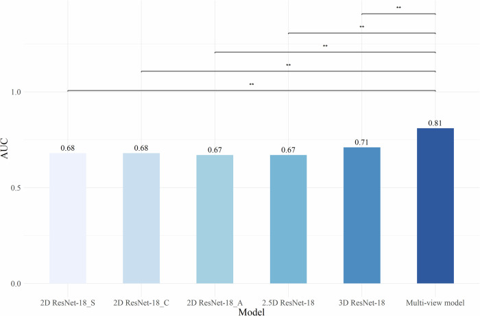
Results: Among all nodules, 18.3% (63) were resolving. The multi-view CNN model achieved an AUC of 0.81, with a mean sensitivity of 0.63 (SD, 0.15) and a mean specificity of 0.93 (SD, 0.02). The model significantly improved performance compared to 2D, 2.5D, or 3D models (p < 0.05). Under the premise of specificity greater than 90% (meaning < 10% of non-resolving nodules are incorrectly identified as resolving), follow-up CT in 14% of individuals could be prevented.
Conclusion: The multi-view CNN model achieved high specificity in discriminating new intermediate nodules that would need follow-up CT by identifying non-resolving nodules. After further validation and optimization, this model may assist with decision-making when new intermediate nodules are found in lung cancer screening.
Critical relevance statement: The multi-view CNN-based model has the potential to reduce unnecessary follow-up scans when new nodules are detected, aiding radiologists in making earlier, more informed decisions.
Key points: Predicting the resolution of new intermediate lung nodules in lung cancer screening CT is a challenge. Our multi-view CNN model showed an AUC of 0.81, a specificity of 0.93, and a sensitivity of 0.63 at the nodule level. The multi-view model demonstrated a significant improvement in AUC compared to the three 2D models, one 2.5D model, and one 3D model.
期刊介绍:
Insights into Imaging (I³) is a peer-reviewed open access journal published under the brand SpringerOpen. All content published in the journal is freely available online to anyone, anywhere!
I³ continuously updates scientific knowledge and progress in best-practice standards in radiology through the publication of original articles and state-of-the-art reviews and opinions, along with recommendations and statements from the leading radiological societies in Europe.
Founded by the European Society of Radiology (ESR), I³ creates a platform for educational material, guidelines and recommendations, and a forum for topics of controversy.
A balanced combination of review articles, original papers, short communications from European radiological congresses and information on society matters makes I³ an indispensable source for current information in this field.
I³ is owned by the ESR, however authors retain copyright to their article according to the Creative Commons Attribution License (see Copyright and License Agreement). All articles can be read, redistributed and reused for free, as long as the author of the original work is cited properly.
The open access fees (article-processing charges) for this journal are kindly sponsored by ESR for all Members.
The journal went open access in 2012, which means that all articles published since then are freely available online.

 求助内容:
求助内容: 应助结果提醒方式:


