Arne Lauer, Luisa Schulte, Artid Skenderi, Nouha Tekiki, Alexander Juerchott, Meysam Sohani, Maurice Ruetters, Franz Sebastian Schwindling, Peter Rammelsberg, Mathias Nittka, Sabine Heiland, Martin Bendszus, Tim Hilgenfeld
{"title":"Dental magnetic resonance imaging for bone loss assessment and disease activity classification in severe periodontitis.","authors":"Arne Lauer, Luisa Schulte, Artid Skenderi, Nouha Tekiki, Alexander Juerchott, Meysam Sohani, Maurice Ruetters, Franz Sebastian Schwindling, Peter Rammelsberg, Mathias Nittka, Sabine Heiland, Martin Bendszus, Tim Hilgenfeld","doi":"10.1186/s13244-025-02004-7","DOIUrl":null,"url":null,"abstract":"<p><strong>Objectives: </strong>To evaluate the reliability and accuracy of dental MRI (dMRI) for volumetric infrabony and furcation bone loss compared to cone-beam computed tomography (CBCT) and to correlate to clinical signs of inflammation in patients with severe periodontitis.</p><p><strong>Methods: </strong>In this cross-sectional study nineteen patients with severe periodontitis underwent standardized clinical examination as well as pre-treatment CBCT and 3T-dMRI. Bone lesion volumetry was performed in CBCT, contrast-enhanced-T1-weighting (T1W + C) and T2-weighting (T2W) dMRI. Lesions whose T2W signal significantly exceeded T1W/CBCT margins (indicating excessive edema) were classified as T2W-mismatch. Volumetric data were compared to clinical findings.</p><p><strong>Results: </strong>Ten female and nine male patients with 253 bony lesions were examined. Reliability for bone lesions was highest in CBCT (ICC [95% CI] T1W + C/T2W/CBCT: 0.78 [0.74-0.83]/0.82 [0.77-0.85]/0.87 [0.94-0.89]). Overall, T1W + C and T2W dMRI strongly correlated with CBCT (r<sub>s</sub> = 0.86 [95% CI: 0.82-0.89], p < 0.001 and r<sub>s</sub> = 0.91 [95% CI: 0.88-0.93], p < 0.001 respectively) but volume was significantly overestimated by dMRI (median percentage error of T1W + C-T2W: 19-55%). A T2W-mismatch was found in 44.1% and correlated with bleeding (85.8% vs. 70.9%, p = 0.005), giving 47.5% sensitivity and 71.2% specificity.</p><p><strong>Conclusions: </strong>While dMRI offers good reliability, T2W- and to a lesser extent T1W + C imaging overestimate infrabony and interradicular periodontal bone lesion volumetry compared to CBCT. While this could increase the risk of overtreatment, dMRI detects periodontal inflammation beyond areas of bone loss, and T2W-mismatch is closely related but not identical to signs of active inflammation in clinical examination. This may provide additional diagnostic information and could serve as a supplemental tool for higher-risk patients.</p><p><strong>Critical relevance statement: </strong>Dental MRI excels in detecting inflammation beyond bone loss, identifying high-risk tissue. This study assesses reliability in evaluating periodontitis-related bone loss, highlighting its tendency to overestimate lesion volume. A novel \"mismatch lesion pattern\" was observed, potentially linked to disease activity.</p><p><strong>Key points: </strong>Dental MRI (dMRI) reliably assesses bone loss in periodontitis but overestimates volume vs. cone-beam computed tomography (CBCT). dMRI detects excess bone marrow edema, indicating inflammation beyond visible bone loss. dMRI could aid periodontal diagnosis and guide targeted therapeutic interventions.</p>","PeriodicalId":13639,"journal":{"name":"Insights into Imaging","volume":"16 1","pages":"134"},"PeriodicalIF":4.5000,"publicationDate":"2025-06-26","publicationTypes":"Journal Article","fieldsOfStudy":null,"isOpenAccess":false,"openAccessPdf":"https://www.ncbi.nlm.nih.gov/pmc/articles/PMC12202246/pdf/","citationCount":"0","resultStr":null,"platform":"Semanticscholar","paperid":null,"PeriodicalName":"Insights into Imaging","FirstCategoryId":"3","ListUrlMain":"https://doi.org/10.1186/s13244-025-02004-7","RegionNum":2,"RegionCategory":"医学","ArticlePicture":[],"TitleCN":null,"AbstractTextCN":null,"PMCID":null,"EPubDate":"","PubModel":"","JCR":"Q1","JCRName":"RADIOLOGY, NUCLEAR MEDICINE & MEDICAL IMAGING","Score":null,"Total":0}
引用次数: 0
Abstract
Objectives: To evaluate the reliability and accuracy of dental MRI (dMRI) for volumetric infrabony and furcation bone loss compared to cone-beam computed tomography (CBCT) and to correlate to clinical signs of inflammation in patients with severe periodontitis.
Methods: In this cross-sectional study nineteen patients with severe periodontitis underwent standardized clinical examination as well as pre-treatment CBCT and 3T-dMRI. Bone lesion volumetry was performed in CBCT, contrast-enhanced-T1-weighting (T1W + C) and T2-weighting (T2W) dMRI. Lesions whose T2W signal significantly exceeded T1W/CBCT margins (indicating excessive edema) were classified as T2W-mismatch. Volumetric data were compared to clinical findings.
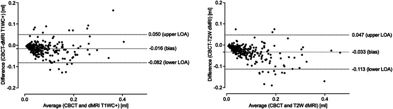
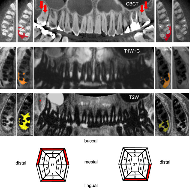
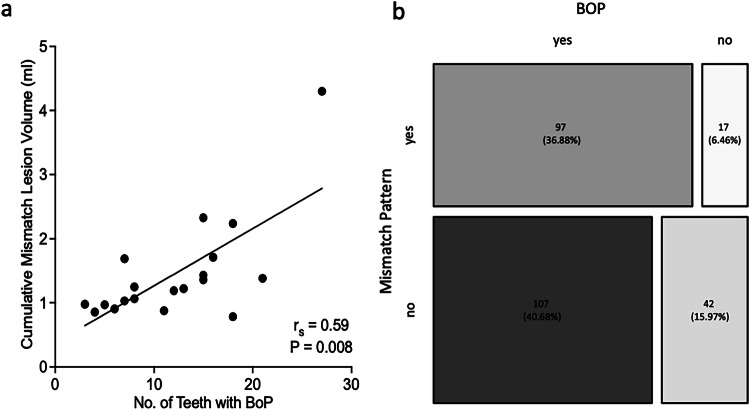
Results: Ten female and nine male patients with 253 bony lesions were examined. Reliability for bone lesions was highest in CBCT (ICC [95% CI] T1W + C/T2W/CBCT: 0.78 [0.74-0.83]/0.82 [0.77-0.85]/0.87 [0.94-0.89]). Overall, T1W + C and T2W dMRI strongly correlated with CBCT (rs = 0.86 [95% CI: 0.82-0.89], p < 0.001 and rs = 0.91 [95% CI: 0.88-0.93], p < 0.001 respectively) but volume was significantly overestimated by dMRI (median percentage error of T1W + C-T2W: 19-55%). A T2W-mismatch was found in 44.1% and correlated with bleeding (85.8% vs. 70.9%, p = 0.005), giving 47.5% sensitivity and 71.2% specificity.
Conclusions: While dMRI offers good reliability, T2W- and to a lesser extent T1W + C imaging overestimate infrabony and interradicular periodontal bone lesion volumetry compared to CBCT. While this could increase the risk of overtreatment, dMRI detects periodontal inflammation beyond areas of bone loss, and T2W-mismatch is closely related but not identical to signs of active inflammation in clinical examination. This may provide additional diagnostic information and could serve as a supplemental tool for higher-risk patients.
Critical relevance statement: Dental MRI excels in detecting inflammation beyond bone loss, identifying high-risk tissue. This study assesses reliability in evaluating periodontitis-related bone loss, highlighting its tendency to overestimate lesion volume. A novel "mismatch lesion pattern" was observed, potentially linked to disease activity.
Key points: Dental MRI (dMRI) reliably assesses bone loss in periodontitis but overestimates volume vs. cone-beam computed tomography (CBCT). dMRI detects excess bone marrow edema, indicating inflammation beyond visible bone loss. dMRI could aid periodontal diagnosis and guide targeted therapeutic interventions.
期刊介绍:
Insights into Imaging (I³) is a peer-reviewed open access journal published under the brand SpringerOpen. All content published in the journal is freely available online to anyone, anywhere!
I³ continuously updates scientific knowledge and progress in best-practice standards in radiology through the publication of original articles and state-of-the-art reviews and opinions, along with recommendations and statements from the leading radiological societies in Europe.
Founded by the European Society of Radiology (ESR), I³ creates a platform for educational material, guidelines and recommendations, and a forum for topics of controversy.
A balanced combination of review articles, original papers, short communications from European radiological congresses and information on society matters makes I³ an indispensable source for current information in this field.
I³ is owned by the ESR, however authors retain copyright to their article according to the Creative Commons Attribution License (see Copyright and License Agreement). All articles can be read, redistributed and reused for free, as long as the author of the original work is cited properly.
The open access fees (article-processing charges) for this journal are kindly sponsored by ESR for all Members.
The journal went open access in 2012, which means that all articles published since then are freely available online.

 求助内容:
求助内容: 应助结果提醒方式:


