Role of Amide Proton Transfer Weighted MRI in Predicting MGMTp Methylation Status, p53-Status, Ki-67 Index, IDH-Status, and ATRX Expression in WHO Grade 4 High Grade Glioma.
IF 2.2 4区 医学Q2 RADIOLOGY, NUCLEAR MEDICINE & MEDICAL IMAGING
Faris Durmo, Jimmy Lätt, Anna Rydelius, Elisabet Englund, Tim Salomonsson, Patrick Liebig, Johan Bengzon, Peter C M van Zijl, Linda Knutsson, Pia C Sundgren
{"title":"Role of Amide Proton Transfer Weighted MRI in Predicting MGMTp Methylation Status, p53-Status, Ki-67 Index, IDH-Status, and ATRX Expression in WHO Grade 4 High Grade Glioma.","authors":"Faris Durmo, Jimmy Lätt, Anna Rydelius, Elisabet Englund, Tim Salomonsson, Patrick Liebig, Johan Bengzon, Peter C M van Zijl, Linda Knutsson, Pia C Sundgren","doi":"10.3390/tomography11060064","DOIUrl":null,"url":null,"abstract":"<p><p><b>Objectives:</b> To assess amide proton transfer weighted (APTw) MR imaging capabilities in differentiating high-grade glial tumors across alpha-thalassemia/mental retardation X-linked (ATRX) expression, tumor-suppressor protein p53 expression (p53), O6-methylguanine-DNA methyltransferase promoter (MGMTp) methylation, isocitrate dehydrogenase (IDH) status, and proliferation marker Ki-67 (Ki-67 index) as a preoperative diagnostic aid. <b>Material & Methods:</b> A total of 42 high-grade glioma WHO grade 4 (HGG) patients were evaluated prospectively (30 males and 12 females). All patients were examined using conventional MRI, including the following: T1w-MPRAGE pre- and post-contrast administration, conventional T2w and 3D FLAIR, and APTw imaging with a 3T MR scanner. Receiver operating characteristic (ROC) curves were calculated for the APTw% mean, median, and max signal for the different molecular biomarkers. A logistic regression model was constructed for combined mean and median APTw% signals for p53 expression. <b>Results:</b> The whole-tumor max APTw% signal could significantly differentiate MGMTp from non-MGMTp HGG, <i>p</i> = 0.035. A cutoff of 4.28% max APTw% signal yielded AUC (area under the curve) = 0.702, with 70.6% sensitivity and 66.7% specificity. The mean/median APTw% signals differed significantly in p53 normal versus p53-overexpressed HGG s: 1.81%/1.83% vs. 1.15%/1.18%, <i>p</i> = 0.002/0.006, respectively. Cutoffs of 1.25%/1.33% for the mean/median APTw% signals yielded AUCs of 0.786/0.757, sensitivities of 76.9%/76.9%, and specificities of 50%/66.2%, <i>p</i> = 0.002/0.006, respectively. A logistic regression model with a combined mean and median APTw% signal for p53 status yielded an AUC = 0.788 and 76.9% sensitivity and 66.2% specificity. ATRX-, IDH- wild type (wt) vs. mutation (mut), and the level of Ki-67 did not differ significantly, but trends were found: IDH-wt and low Ki-67 showed higher mean/median/max APTw% signals vs. IDH-mut and high Ki-67, respectively. ATRX-wt vs. mutation showed higher mean and median APTw% signals but lower max APTw% signal. <b>Conclusions</b>: APTw imaging can potentially be a useful marker for the stratification of p53 expression and MGMT status in high-grade glioma in the preoperative setting and potentially aid surgical decision-making.</p>","PeriodicalId":51330,"journal":{"name":"Tomography","volume":"11 6","pages":""},"PeriodicalIF":2.2000,"publicationDate":"2025-05-31","publicationTypes":"Journal Article","fieldsOfStudy":null,"isOpenAccess":false,"openAccessPdf":"https://www.ncbi.nlm.nih.gov/pmc/articles/PMC12196788/pdf/","citationCount":"0","resultStr":null,"platform":"Semanticscholar","paperid":null,"PeriodicalName":"Tomography","FirstCategoryId":"3","ListUrlMain":"https://doi.org/10.3390/tomography11060064","RegionNum":4,"RegionCategory":"医学","ArticlePicture":[],"TitleCN":null,"AbstractTextCN":null,"PMCID":null,"EPubDate":"","PubModel":"","JCR":"Q2","JCRName":"RADIOLOGY, NUCLEAR MEDICINE & MEDICAL IMAGING","Score":null,"Total":0}
引用次数: 0
Abstract
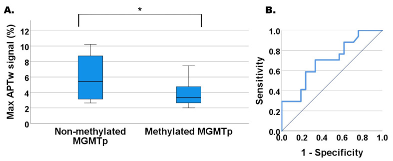
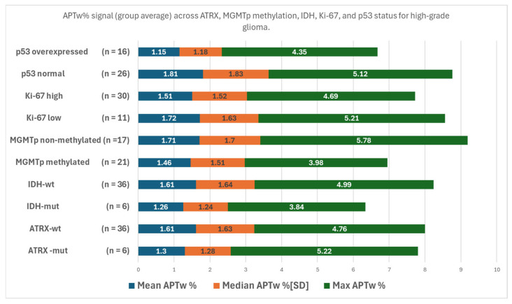
Objectives: To assess amide proton transfer weighted (APTw) MR imaging capabilities in differentiating high-grade glial tumors across alpha-thalassemia/mental retardation X-linked (ATRX) expression, tumor-suppressor protein p53 expression (p53), O6-methylguanine-DNA methyltransferase promoter (MGMTp) methylation, isocitrate dehydrogenase (IDH) status, and proliferation marker Ki-67 (Ki-67 index) as a preoperative diagnostic aid. Material & Methods: A total of 42 high-grade glioma WHO grade 4 (HGG) patients were evaluated prospectively (30 males and 12 females). All patients were examined using conventional MRI, including the following: T1w-MPRAGE pre- and post-contrast administration, conventional T2w and 3D FLAIR, and APTw imaging with a 3T MR scanner. Receiver operating characteristic (ROC) curves were calculated for the APTw% mean, median, and max signal for the different molecular biomarkers. A logistic regression model was constructed for combined mean and median APTw% signals for p53 expression. Results: The whole-tumor max APTw% signal could significantly differentiate MGMTp from non-MGMTp HGG, p = 0.035. A cutoff of 4.28% max APTw% signal yielded AUC (area under the curve) = 0.702, with 70.6% sensitivity and 66.7% specificity. The mean/median APTw% signals differed significantly in p53 normal versus p53-overexpressed HGG s: 1.81%/1.83% vs. 1.15%/1.18%, p = 0.002/0.006, respectively. Cutoffs of 1.25%/1.33% for the mean/median APTw% signals yielded AUCs of 0.786/0.757, sensitivities of 76.9%/76.9%, and specificities of 50%/66.2%, p = 0.002/0.006, respectively. A logistic regression model with a combined mean and median APTw% signal for p53 status yielded an AUC = 0.788 and 76.9% sensitivity and 66.2% specificity. ATRX-, IDH- wild type (wt) vs. mutation (mut), and the level of Ki-67 did not differ significantly, but trends were found: IDH-wt and low Ki-67 showed higher mean/median/max APTw% signals vs. IDH-mut and high Ki-67, respectively. ATRX-wt vs. mutation showed higher mean and median APTw% signals but lower max APTw% signal. Conclusions: APTw imaging can potentially be a useful marker for the stratification of p53 expression and MGMT status in high-grade glioma in the preoperative setting and potentially aid surgical decision-making.
TomographyMedicine-Radiology, Nuclear Medicine and Imaging
CiteScore
2.70
自引率
10.50%
发文量
222
期刊介绍:
TomographyTM publishes basic (technical and pre-clinical) and clinical scientific articles which involve the advancement of imaging technologies. Tomography encompasses studies that use single or multiple imaging modalities including for example CT, US, PET, SPECT, MR and hyperpolarization technologies, as well as optical modalities (i.e. bioluminescence, photoacoustic, endomicroscopy, fiber optic imaging and optical computed tomography) in basic sciences, engineering, preclinical and clinical medicine.
Tomography also welcomes studies involving exploration and refinement of contrast mechanisms and image-derived metrics within and across modalities toward the development of novel imaging probes for image-based feedback and intervention. The use of imaging in biology and medicine provides unparalleled opportunities to noninvasively interrogate tissues to obtain real-time dynamic and quantitative information required for diagnosis and response to interventions and to follow evolving pathological conditions. As multi-modal studies and the complexities of imaging technologies themselves are ever increasing to provide advanced information to scientists and clinicians.
Tomography provides a unique publication venue allowing investigators the opportunity to more precisely communicate integrated findings related to the diverse and heterogeneous features associated with underlying anatomical, physiological, functional, metabolic and molecular genetic activities of normal and diseased tissue. Thus Tomography publishes peer-reviewed articles which involve the broad use of imaging of any tissue and disease type including both preclinical and clinical investigations. In addition, hardware/software along with chemical and molecular probe advances are welcome as they are deemed to significantly contribute towards the long-term goal of improving the overall impact of imaging on scientific and clinical discovery.

 求助内容:
求助内容: 应助结果提醒方式:


