Abinand C Rejimon, Anuradha G Trivedi, Vicki Huang, Karthik K Ramesh, Natia Esiashvilli, Eduard Schreibmann, Hyunsuk Shim, Kartik Reddy, Bree R Eaton
{"title":"Longitudinal Overlap and Metabolite Analysis in Spectroscopic MRI-Guided Proton Beam Therapy in Pediatric High-Grade Glioma.","authors":"Abinand C Rejimon, Anuradha G Trivedi, Vicki Huang, Karthik K Ramesh, Natia Esiashvilli, Eduard Schreibmann, Hyunsuk Shim, Kartik Reddy, Bree R Eaton","doi":"10.3390/tomography11060071","DOIUrl":null,"url":null,"abstract":"<p><strong>Background: </strong>Pediatric high-grade glioma (pHGG) is a highly aggressive cancer with unique biology distinct from adult high-grade glioma, limiting the effectiveness of standard treatment protocols derived from adult research.</p><p><strong>Objective: </strong>The purpose of this report is to present preliminary results from an ongoing pilot study integrating spectroscopic magnetic resonance imaging (sMRI) to guide proton beam therapy and longitudinal imaging analysis in pediatric patients with high-grade glioma (pHGG).</p><p><strong>Methods: </strong>Thirteen pediatric patients under 21 years old with supratentorial WHO grade III-IV glioma underwent baseline and serial whole-brain spectroscopic MRI alongside standard structural MRIs. Radiation targets were defined using T1-weighted contrast enhanced, T2-FLAIR, and Cho/NAA ≥ 2X maps. Longitudinal analyses included voxel-level metabolic change maps and spatial overlap metrics comparing pre-proton therapy and post-.</p><p><strong>Results: </strong>Six patients had sufficient longitudinal data; five received sMRI-guided PBT. Significant positive correlation (R<sup>2</sup> = 0.89, <i>p</i> < 0.0001) was observed between T2-FLAIR and Cho/NAA ≥ 2X volumes. Voxel-level difference maps of Cho/NAA and Choline revealed dynamic metabolic changes across follow-up scans. Analyzing Cho/NAA and Cho changes over time allowed differentiation between true progression and pseudoprogression, which conventional MRI alone struggles to achieve.</p><p><strong>Conclusions: </strong>Longitudinal sMRI enhanced metabolic tracking in pHGG, detects early tumor changes, and refines RT targeting beyond structural imaging. This first in-kind study highlights the potential of sMRI biomarkers in tracking treatment effects and emphasizes the complementary roles of metabolic and radiographic metrics in evaluating therapy response in pHGG.</p>","PeriodicalId":51330,"journal":{"name":"Tomography","volume":"11 6","pages":""},"PeriodicalIF":2.2000,"publicationDate":"2025-06-19","publicationTypes":"Journal Article","fieldsOfStudy":null,"isOpenAccess":false,"openAccessPdf":"https://www.ncbi.nlm.nih.gov/pmc/articles/PMC12196607/pdf/","citationCount":"0","resultStr":null,"platform":"Semanticscholar","paperid":null,"PeriodicalName":"Tomography","FirstCategoryId":"3","ListUrlMain":"https://doi.org/10.3390/tomography11060071","RegionNum":4,"RegionCategory":"医学","ArticlePicture":[],"TitleCN":null,"AbstractTextCN":null,"PMCID":null,"EPubDate":"","PubModel":"","JCR":"Q2","JCRName":"RADIOLOGY, NUCLEAR MEDICINE & MEDICAL IMAGING","Score":null,"Total":0}
引用次数: 0
Abstract
Background: Pediatric high-grade glioma (pHGG) is a highly aggressive cancer with unique biology distinct from adult high-grade glioma, limiting the effectiveness of standard treatment protocols derived from adult research.
Objective: The purpose of this report is to present preliminary results from an ongoing pilot study integrating spectroscopic magnetic resonance imaging (sMRI) to guide proton beam therapy and longitudinal imaging analysis in pediatric patients with high-grade glioma (pHGG).
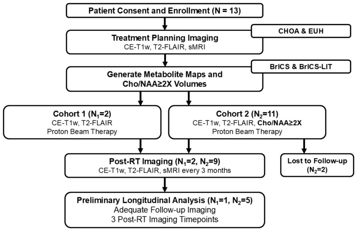
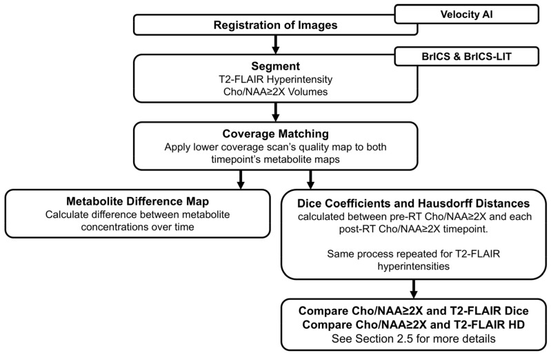
Methods: Thirteen pediatric patients under 21 years old with supratentorial WHO grade III-IV glioma underwent baseline and serial whole-brain spectroscopic MRI alongside standard structural MRIs. Radiation targets were defined using T1-weighted contrast enhanced, T2-FLAIR, and Cho/NAA ≥ 2X maps. Longitudinal analyses included voxel-level metabolic change maps and spatial overlap metrics comparing pre-proton therapy and post-.
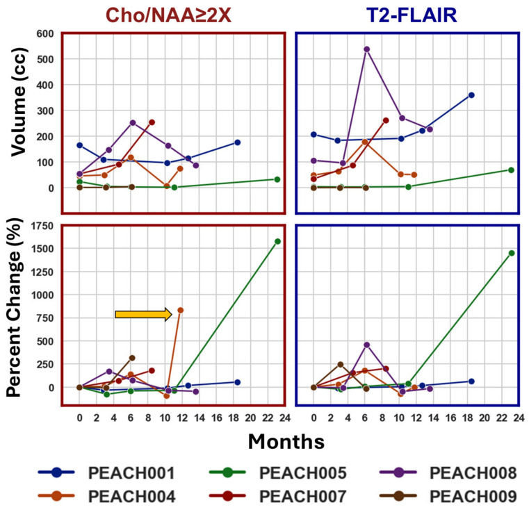
Results: Six patients had sufficient longitudinal data; five received sMRI-guided PBT. Significant positive correlation (R2 = 0.89, p < 0.0001) was observed between T2-FLAIR and Cho/NAA ≥ 2X volumes. Voxel-level difference maps of Cho/NAA and Choline revealed dynamic metabolic changes across follow-up scans. Analyzing Cho/NAA and Cho changes over time allowed differentiation between true progression and pseudoprogression, which conventional MRI alone struggles to achieve.
Conclusions: Longitudinal sMRI enhanced metabolic tracking in pHGG, detects early tumor changes, and refines RT targeting beyond structural imaging. This first in-kind study highlights the potential of sMRI biomarkers in tracking treatment effects and emphasizes the complementary roles of metabolic and radiographic metrics in evaluating therapy response in pHGG.
TomographyMedicine-Radiology, Nuclear Medicine and Imaging
CiteScore
2.70
自引率
10.50%
发文量
222
期刊介绍:
TomographyTM publishes basic (technical and pre-clinical) and clinical scientific articles which involve the advancement of imaging technologies. Tomography encompasses studies that use single or multiple imaging modalities including for example CT, US, PET, SPECT, MR and hyperpolarization technologies, as well as optical modalities (i.e. bioluminescence, photoacoustic, endomicroscopy, fiber optic imaging and optical computed tomography) in basic sciences, engineering, preclinical and clinical medicine.
Tomography also welcomes studies involving exploration and refinement of contrast mechanisms and image-derived metrics within and across modalities toward the development of novel imaging probes for image-based feedback and intervention. The use of imaging in biology and medicine provides unparalleled opportunities to noninvasively interrogate tissues to obtain real-time dynamic and quantitative information required for diagnosis and response to interventions and to follow evolving pathological conditions. As multi-modal studies and the complexities of imaging technologies themselves are ever increasing to provide advanced information to scientists and clinicians.
Tomography provides a unique publication venue allowing investigators the opportunity to more precisely communicate integrated findings related to the diverse and heterogeneous features associated with underlying anatomical, physiological, functional, metabolic and molecular genetic activities of normal and diseased tissue. Thus Tomography publishes peer-reviewed articles which involve the broad use of imaging of any tissue and disease type including both preclinical and clinical investigations. In addition, hardware/software along with chemical and molecular probe advances are welcome as they are deemed to significantly contribute towards the long-term goal of improving the overall impact of imaging on scientific and clinical discovery.

 求助内容:
求助内容: 应助结果提醒方式:


