Elif Yıldızer, Saliha Kubra Sari, Fatih Peker, Ali Riza Erdogan, Kevser Sancak, Sinan Yasin Ertem
{"title":"Assessment of Mandibular Bone Architecture in Patients with Endocrine Disorders Using Fractal Dimension and Histogram Analysis.","authors":"Elif Yıldızer, Saliha Kubra Sari, Fatih Peker, Ali Riza Erdogan, Kevser Sancak, Sinan Yasin Ertem","doi":"10.3390/tomography11060070","DOIUrl":null,"url":null,"abstract":"<p><strong>Objective: </strong>Endocrine disorders, including diabetes mellitus and thyroid dysfunctions, can significantly impact bone metabolism and structure. This study aimed to assess mandibular trabecular architecture in patients with type 1 diabetes mellitus (T1DM), type 2 diabetes mellitus (T2DM), hyperthyroidism, and hypothyroidism using fractal dimension (FD) and histogram analyses (HA), comparing the findings with a healthy control group.</p><p><strong>Methods: </strong>This retrospective study analyzed panoramic radiographs from 200 individuals, comprising 40 patients in each of the four endocrine disorder groups and 40 healthy controls. Fractal dimension and histogram-based pixel intensity analyses were conducted using ImageJ™ (version 1.53) software. Four standardized regions of interest (ROI) were evaluated on the right mandible, and statistical comparisons were conducted across groups using one-way analysis of variance (ANOVA), <i>t</i>-test, Mann-Whitney U, and Spearman correlation analyses.</p><p><strong>Results: </strong>Age and gender distributions did not differ significantly between groups. FD analysis revealed a significant reduction at ROI1 in the hyperthyroidism group compared to controls (<i>p</i> = 0.018); however, no other significant FD differences were observed among the remaining groups or ROIs. A significant positive correlation was found between FD and histogram values at ROI1 and ROI2 (<i>p</i> < 0.001), while pixel intensity values did not differ significantly across groups in any ROI.</p><p><strong>Conclusion: </strong>Although no significant differences were found in diabetic groups, the decreased FD in hyperthyroid patients suggests that FD analysis may be a useful non-invasive method to detect subtle bone alterations. Further research with larger sample sizes and comprehensive biochemical analyses are needed to confirm these findings.</p>","PeriodicalId":51330,"journal":{"name":"Tomography","volume":"11 6","pages":""},"PeriodicalIF":2.2000,"publicationDate":"2025-06-18","publicationTypes":"Journal Article","fieldsOfStudy":null,"isOpenAccess":false,"openAccessPdf":"https://www.ncbi.nlm.nih.gov/pmc/articles/PMC12196900/pdf/","citationCount":"0","resultStr":null,"platform":"Semanticscholar","paperid":null,"PeriodicalName":"Tomography","FirstCategoryId":"3","ListUrlMain":"https://doi.org/10.3390/tomography11060070","RegionNum":4,"RegionCategory":"医学","ArticlePicture":[],"TitleCN":null,"AbstractTextCN":null,"PMCID":null,"EPubDate":"","PubModel":"","JCR":"Q2","JCRName":"RADIOLOGY, NUCLEAR MEDICINE & MEDICAL IMAGING","Score":null,"Total":0}
引用次数: 0
Abstract
Objective: Endocrine disorders, including diabetes mellitus and thyroid dysfunctions, can significantly impact bone metabolism and structure. This study aimed to assess mandibular trabecular architecture in patients with type 1 diabetes mellitus (T1DM), type 2 diabetes mellitus (T2DM), hyperthyroidism, and hypothyroidism using fractal dimension (FD) and histogram analyses (HA), comparing the findings with a healthy control group.
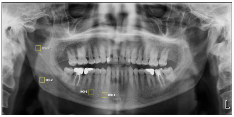
Methods: This retrospective study analyzed panoramic radiographs from 200 individuals, comprising 40 patients in each of the four endocrine disorder groups and 40 healthy controls. Fractal dimension and histogram-based pixel intensity analyses were conducted using ImageJ™ (version 1.53) software. Four standardized regions of interest (ROI) were evaluated on the right mandible, and statistical comparisons were conducted across groups using one-way analysis of variance (ANOVA), t-test, Mann-Whitney U, and Spearman correlation analyses.
Results: Age and gender distributions did not differ significantly between groups. FD analysis revealed a significant reduction at ROI1 in the hyperthyroidism group compared to controls (p = 0.018); however, no other significant FD differences were observed among the remaining groups or ROIs. A significant positive correlation was found between FD and histogram values at ROI1 and ROI2 (p < 0.001), while pixel intensity values did not differ significantly across groups in any ROI.
Conclusion: Although no significant differences were found in diabetic groups, the decreased FD in hyperthyroid patients suggests that FD analysis may be a useful non-invasive method to detect subtle bone alterations. Further research with larger sample sizes and comprehensive biochemical analyses are needed to confirm these findings.
TomographyMedicine-Radiology, Nuclear Medicine and Imaging
CiteScore
2.70
自引率
10.50%
发文量
222
期刊介绍:
TomographyTM publishes basic (technical and pre-clinical) and clinical scientific articles which involve the advancement of imaging technologies. Tomography encompasses studies that use single or multiple imaging modalities including for example CT, US, PET, SPECT, MR and hyperpolarization technologies, as well as optical modalities (i.e. bioluminescence, photoacoustic, endomicroscopy, fiber optic imaging and optical computed tomography) in basic sciences, engineering, preclinical and clinical medicine.
Tomography also welcomes studies involving exploration and refinement of contrast mechanisms and image-derived metrics within and across modalities toward the development of novel imaging probes for image-based feedback and intervention. The use of imaging in biology and medicine provides unparalleled opportunities to noninvasively interrogate tissues to obtain real-time dynamic and quantitative information required for diagnosis and response to interventions and to follow evolving pathological conditions. As multi-modal studies and the complexities of imaging technologies themselves are ever increasing to provide advanced information to scientists and clinicians.
Tomography provides a unique publication venue allowing investigators the opportunity to more precisely communicate integrated findings related to the diverse and heterogeneous features associated with underlying anatomical, physiological, functional, metabolic and molecular genetic activities of normal and diseased tissue. Thus Tomography publishes peer-reviewed articles which involve the broad use of imaging of any tissue and disease type including both preclinical and clinical investigations. In addition, hardware/software along with chemical and molecular probe advances are welcome as they are deemed to significantly contribute towards the long-term goal of improving the overall impact of imaging on scientific and clinical discovery.

 求助内容:
求助内容: 应助结果提醒方式:


