{"title":"Differentiating Malignant and Healthy Areas in Isolated Kidney Samples Through Infrared Visualization Techniques.","authors":"Besarion Partsvania, Tamaz Sulaberidze, Alexandre Khuskivadze, Sophio Abazadze, Teimuraz Gogoladze, Nutsa Khuskivadze","doi":"10.14740/wjon2593","DOIUrl":null,"url":null,"abstract":"<p><strong>Background: </strong>Because partial nephrectomy (PN) may remove malignant tissue while maintaining kidney function, it is currently the gold standard for nephrectomy. However, the blood arteries that supply the kidney are clamped at the start of the procedure. The most common method for evaluating surgical margins during PN is intraoperative frozen section (FS) evaluation. Its long duration and high false-negative rate question its reliability and efficacy. This encouraged us to search for a much quicker and easier method.</p><p><strong>Methods: </strong>The infrared (IR) imaging approach uses the differences in optical density between tumor and healthy tissue to create the sharp contrast in the IR images. The cancerous kidneys were examined after a radical nephrectomy. Following the removal of the cancerous tissue and some of the surrounding healthy tissue, the samples were examined using the IR method. For the IR analysis, we created specific software. Following that, tissue samples taken from both healthy and malignant areas were subjected to a histomorphological analysis.</p><p><strong>Results: </strong>Experiments showed that malignant tissue appeared as areas of high blackness in the IR picture, while healthy tissue appeared as areas of high illumination. Our software highlighted the areas of the IR image that were associated with the healthy and malignant portions, computed their average brightness, and calculated the ratio of the average illumination (RAI) of the malignant area to that of the healthy area. RAI is an interval of numbers obtained as a result of dividing the average brightness of all dark areas in all examined samples by all light areas of all examined samples. The 95% probability interval for RAIs taking place, which ranged from 0.25 to 0.41, was calculated. The location of the malignancy was then identified by a histomorphological examination. The compliance between histomorphological results and the outcomes of IR examination was confirmed in all cases.</p><p><strong>Conclusions: </strong>The IR imaging technique offers significant promise for improving the accuracy and efficiency of margin assessment during kidney cancer surgeries. The IR imaging technique can provide immediate feedback on the tumor boundaries, which could potentially reduce the duration of warm ischemia during surgery. Subsequent investigations should be focused on verifying the technology in further clinical trials and investigating its integration into the surgical process, which could result in its acceptance as a standard instrument for intraoperative decision-making in kidney cancer operations.</p>","PeriodicalId":46797,"journal":{"name":"World Journal of Oncology","volume":"16 3","pages":"311-316"},"PeriodicalIF":2.2000,"publicationDate":"2025-06-01","publicationTypes":"Journal Article","fieldsOfStudy":null,"isOpenAccess":false,"openAccessPdf":"https://www.ncbi.nlm.nih.gov/pmc/articles/PMC12185124/pdf/","citationCount":"0","resultStr":null,"platform":"Semanticscholar","paperid":null,"PeriodicalName":"World Journal of Oncology","FirstCategoryId":"1085","ListUrlMain":"https://doi.org/10.14740/wjon2593","RegionNum":0,"RegionCategory":null,"ArticlePicture":[],"TitleCN":null,"AbstractTextCN":null,"PMCID":null,"EPubDate":"2025/6/14 0:00:00","PubModel":"Epub","JCR":"Q3","JCRName":"ONCOLOGY","Score":null,"Total":0}
引用次数: 0
Abstract
Background: Because partial nephrectomy (PN) may remove malignant tissue while maintaining kidney function, it is currently the gold standard for nephrectomy. However, the blood arteries that supply the kidney are clamped at the start of the procedure. The most common method for evaluating surgical margins during PN is intraoperative frozen section (FS) evaluation. Its long duration and high false-negative rate question its reliability and efficacy. This encouraged us to search for a much quicker and easier method.
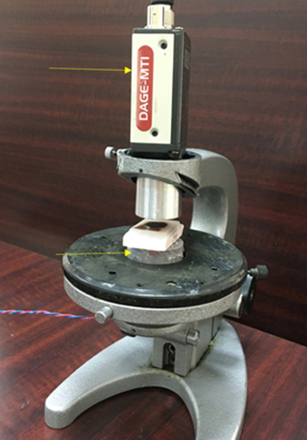
Methods: The infrared (IR) imaging approach uses the differences in optical density between tumor and healthy tissue to create the sharp contrast in the IR images. The cancerous kidneys were examined after a radical nephrectomy. Following the removal of the cancerous tissue and some of the surrounding healthy tissue, the samples were examined using the IR method. For the IR analysis, we created specific software. Following that, tissue samples taken from both healthy and malignant areas were subjected to a histomorphological analysis.
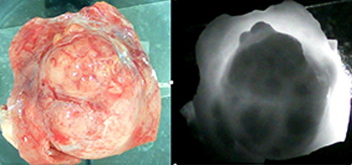
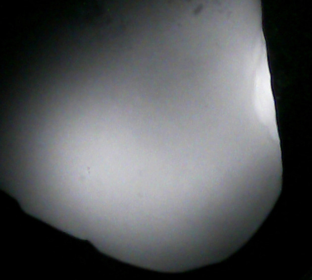
Results: Experiments showed that malignant tissue appeared as areas of high blackness in the IR picture, while healthy tissue appeared as areas of high illumination. Our software highlighted the areas of the IR image that were associated with the healthy and malignant portions, computed their average brightness, and calculated the ratio of the average illumination (RAI) of the malignant area to that of the healthy area. RAI is an interval of numbers obtained as a result of dividing the average brightness of all dark areas in all examined samples by all light areas of all examined samples. The 95% probability interval for RAIs taking place, which ranged from 0.25 to 0.41, was calculated. The location of the malignancy was then identified by a histomorphological examination. The compliance between histomorphological results and the outcomes of IR examination was confirmed in all cases.
Conclusions: The IR imaging technique offers significant promise for improving the accuracy and efficiency of margin assessment during kidney cancer surgeries. The IR imaging technique can provide immediate feedback on the tumor boundaries, which could potentially reduce the duration of warm ischemia during surgery. Subsequent investigations should be focused on verifying the technology in further clinical trials and investigating its integration into the surgical process, which could result in its acceptance as a standard instrument for intraoperative decision-making in kidney cancer operations.
期刊介绍:
World Journal of Oncology, bimonthly, publishes original contributions describing basic research and clinical investigation of cancer, on the cellular, molecular, prevention, diagnosis, therapy and prognosis aspects. The submissions can be basic research or clinical investigation oriented. This journal welcomes those submissions focused on the clinical trials of new treatment modalities for cancer, and those submissions focused on molecular or cellular research of the oncology pathogenesis. Case reports submitted for consideration of publication should explore either a novel genomic event/description or a new safety signal from an oncolytic agent. The areas of interested manuscripts are these disciplines: tumor immunology and immunotherapy; cancer molecular pharmacology and chemotherapy; drug sensitivity and resistance; cancer epidemiology; clinical trials; cancer pathology; radiobiology and radiation oncology; solid tumor oncology; hematological malignancies; surgical oncology; pediatric oncology; molecular oncology and cancer genes; gene therapy; cancer endocrinology; cancer metastasis; prevention and diagnosis of cancer; other cancer related subjects. The types of manuscripts accepted are original article, review, editorial, short communication, case report, letter to the editor, book review.
 求助内容:
求助内容: 应助结果提醒方式:


