Mika Naganawa, Ming-Qiang Zheng, Jean-Dominique Gallezot, Robin Bonomi, Jiwei Gu, Hong Gao, Swanee Jacutin-Porte, Nabeel B Nabulsi, Michel Koole, Koen Van Laere, David Matuskey, Yiyun Huang, Richard E Carson
{"title":"Assessment of test-retest reproducibility by [<sup>18</sup>F]Bavarostat for PET imaging of HDAC6.","authors":"Mika Naganawa, Ming-Qiang Zheng, Jean-Dominique Gallezot, Robin Bonomi, Jiwei Gu, Hong Gao, Swanee Jacutin-Porte, Nabeel B Nabulsi, Michel Koole, Koen Van Laere, David Matuskey, Yiyun Huang, Richard E Carson","doi":"10.1186/s13550-025-01268-w","DOIUrl":null,"url":null,"abstract":"<p><strong>Background: </strong>Histone deacetylase 6 (HDAC6) is an enzyme pivotal for gene regulation, influencing cellular pathways through protein deacetylation. HDAC6 is a potential therapeutic target in diseases such as cancer and neurodegenerative disorders. Koole et al. investigated brain binding of [<sup>18</sup>F]Bavarostat, an HDAC6 inhibitor, in healthy participants, revealing an absolute test-retest variability (aTRV) of 7.7% (n = 4) for the distribution volume (V<sub>T</sub>) with a 1-day interscan interval. This study aims to evaluate test-retest reproducibility with a more extended interscan interval.</p><p><strong>Results: </strong>Six participants (3 M/3F) underwent a test-retest scan, each lasting for 120 min using a 4-ring Biograph mCT PET/CT scanner. Arterial blood sampling and metabolite analysis were performed to derive the input function. The two scans were 28 ± 12 days apart (14-43 days, n = 6). Regional time-activity curves (TACs) were generated for 15 regions of interest (ROIs). Kinetic analysis of the 120-min TACs was performed using one-tissue and two-tissue compartment models (1TC, 2TC) and multilinear analysis-1 (MA1) to quantify V<sub>T</sub> values and compute absolute test-retest variability (aTRV). The effects of scan duration (60 to 120 min) and MA1 t* setting on aTRV and bias were investigated. Careful analysis of the plasma HPLC data was needed since metabolites eluted close in time to the parent. The MA1 model (t* = 40 min) adequately described regional TACs and produced stable kinetic parameters with good agreement to 2TC (MA1 V<sub>T</sub>=0.98 × 2TC V<sub>T</sub> + 0.48, bias: -0.1%) while 1TC underestimated V<sub>T</sub> by 5.1%. Regional V<sub>T</sub> values exhibited a relatively uniform pattern, highest in the amygdala and lowest in the centrum semiovale. Individual aTRV values ranged from 2 to 9%. Scan durations between 100 and 120 min provided the most consistent results, with minimal bias and acceptable aTRV across all tested t* values. Although a 90-minute scan with t*=10 or 20-minute balanced scan time and aTRV, optimal parameters varied by brain region. Smaller regions (e.g., amygdala) required longer scans to achieve reliable V<sub>T</sub> quantification.</p><p><strong>Conclusions: </strong>The test-retest variability of [<sup>18</sup>F]Bavarostat V<sub>T</sub> values demonstrated favorable results for a one-month scan interval, comparable to the reported values.</p>","PeriodicalId":11611,"journal":{"name":"EJNMMI Research","volume":"15 1","pages":"76"},"PeriodicalIF":3.1000,"publicationDate":"2025-06-21","publicationTypes":"Journal Article","fieldsOfStudy":null,"isOpenAccess":false,"openAccessPdf":"https://www.ncbi.nlm.nih.gov/pmc/articles/PMC12182542/pdf/","citationCount":"0","resultStr":null,"platform":"Semanticscholar","paperid":null,"PeriodicalName":"EJNMMI Research","FirstCategoryId":"3","ListUrlMain":"https://doi.org/10.1186/s13550-025-01268-w","RegionNum":3,"RegionCategory":"医学","ArticlePicture":[],"TitleCN":null,"AbstractTextCN":null,"PMCID":null,"EPubDate":"","PubModel":"","JCR":"Q1","JCRName":"RADIOLOGY, NUCLEAR MEDICINE & MEDICAL IMAGING","Score":null,"Total":0}
引用次数: 0
Abstract
Background: Histone deacetylase 6 (HDAC6) is an enzyme pivotal for gene regulation, influencing cellular pathways through protein deacetylation. HDAC6 is a potential therapeutic target in diseases such as cancer and neurodegenerative disorders. Koole et al. investigated brain binding of [18F]Bavarostat, an HDAC6 inhibitor, in healthy participants, revealing an absolute test-retest variability (aTRV) of 7.7% (n = 4) for the distribution volume (VT) with a 1-day interscan interval. This study aims to evaluate test-retest reproducibility with a more extended interscan interval.
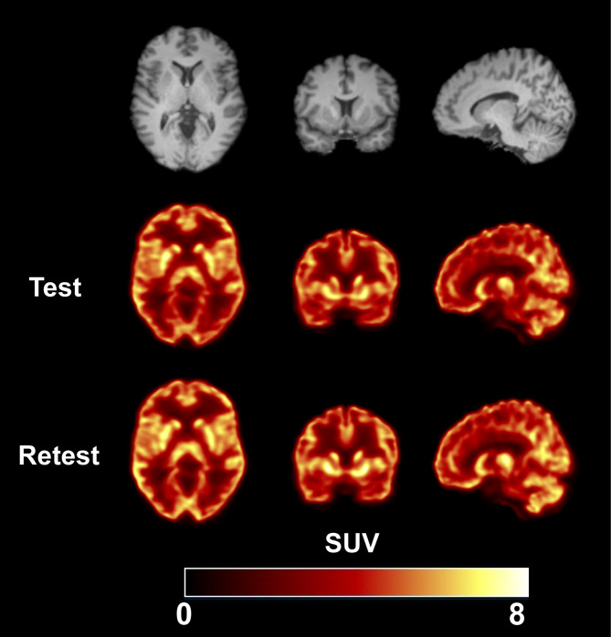
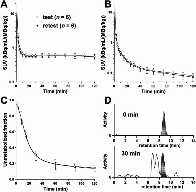
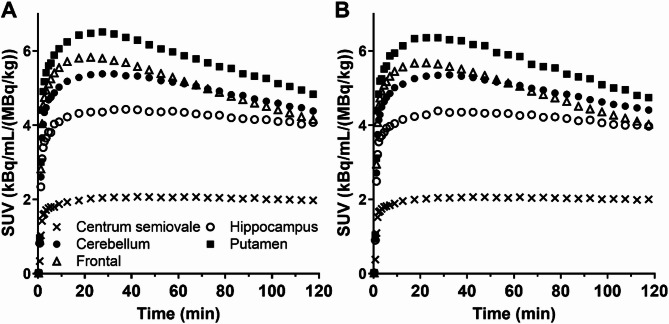
Results: Six participants (3 M/3F) underwent a test-retest scan, each lasting for 120 min using a 4-ring Biograph mCT PET/CT scanner. Arterial blood sampling and metabolite analysis were performed to derive the input function. The two scans were 28 ± 12 days apart (14-43 days, n = 6). Regional time-activity curves (TACs) were generated for 15 regions of interest (ROIs). Kinetic analysis of the 120-min TACs was performed using one-tissue and two-tissue compartment models (1TC, 2TC) and multilinear analysis-1 (MA1) to quantify VT values and compute absolute test-retest variability (aTRV). The effects of scan duration (60 to 120 min) and MA1 t* setting on aTRV and bias were investigated. Careful analysis of the plasma HPLC data was needed since metabolites eluted close in time to the parent. The MA1 model (t* = 40 min) adequately described regional TACs and produced stable kinetic parameters with good agreement to 2TC (MA1 VT=0.98 × 2TC VT + 0.48, bias: -0.1%) while 1TC underestimated VT by 5.1%. Regional VT values exhibited a relatively uniform pattern, highest in the amygdala and lowest in the centrum semiovale. Individual aTRV values ranged from 2 to 9%. Scan durations between 100 and 120 min provided the most consistent results, with minimal bias and acceptable aTRV across all tested t* values. Although a 90-minute scan with t*=10 or 20-minute balanced scan time and aTRV, optimal parameters varied by brain region. Smaller regions (e.g., amygdala) required longer scans to achieve reliable VT quantification.
Conclusions: The test-retest variability of [18F]Bavarostat VT values demonstrated favorable results for a one-month scan interval, comparable to the reported values.
EJNMMI ResearchRADIOLOGY, NUCLEAR MEDICINE & MEDICAL IMAGING&nb-
CiteScore
5.90
自引率
3.10%
发文量
72
审稿时长
13 weeks
期刊介绍:
EJNMMI Research publishes new basic, translational and clinical research in the field of nuclear medicine and molecular imaging. Regular features include original research articles, rapid communication of preliminary data on innovative research, interesting case reports, editorials, and letters to the editor. Educational articles on basic sciences, fundamental aspects and controversy related to pre-clinical and clinical research or ethical aspects of research are also welcome. Timely reviews provide updates on current applications, issues in imaging research and translational aspects of nuclear medicine and molecular imaging technologies.
The main emphasis is placed on the development of targeted imaging with radiopharmaceuticals within the broader context of molecular probes to enhance understanding and characterisation of the complex biological processes underlying disease and to develop, test and guide new treatment modalities, including radionuclide therapy.

 求助内容:
求助内容: 应助结果提醒方式:


