{"title":"Advances of MR imaging in glioma: what the neurosurgeon needs to know.","authors":"Anna Falk Delgado","doi":"10.1007/s00701-025-06593-6","DOIUrl":null,"url":null,"abstract":"<p><p>Glial tumors and especially glioblastoma present a major challenge in neuro-oncology due to their infiltrative growth, resistance to therapy, and poor overall survival-despite aggressive treatments such as maximal safe resection and chemoradiotherapy. These tumors typically manifest through neurological symptoms such as seizures, headaches, and signs of increased intracranial pressure, prompting urgent neuroimaging. At initial diagnosis, MRI plays a central role in differentiating true neoplasms from tumor mimics, including inflammatory or infectious conditions. Advanced techniques such as perfusion-weighted imaging (PWI) and diffusion-weighted imaging (DWI) enhance diagnostic specificity and may prevent unnecessary surgical intervention. In the preoperative phase, MRI contributes to surgical planning through the use of functional MRI (fMRI) and diffusion tensor imaging (DTI), enabling localization of eloquent cortex and white matter tracts. These modalities support safer resections by informing trajectory planning and risk assessment. Emerging MR techniques, including magnetic resonance spectroscopy, amide proton transfer imaging, and 2HG quantification, offer further potential in delineating tumor infiltration beyond contrast-enhancing margins. Postoperatively, MRI is important for evaluating residual tumor, detecting surgical complications, and guiding radiotherapy planning. During treatment surveillance, MRI assists in distinguishing true progression from pseudoprogression or radiation necrosis, thereby guiding decisions on additional surgery, changes in systemic therapy, or inclusion into clinical trials. The continued evolution of MRI hardware, software, and image analysis-particularly with the integration of machine learning-will be critical for supporting precision neurosurgical oncology. This review highlights how advanced MRI techniques can inform clinical decision-making at each stage of care in patients with high-grade gliomas.</p>","PeriodicalId":7370,"journal":{"name":"Acta Neurochirurgica","volume":"167 1","pages":"174"},"PeriodicalIF":1.9000,"publicationDate":"2025-06-21","publicationTypes":"Journal Article","fieldsOfStudy":null,"isOpenAccess":false,"openAccessPdf":"https://www.ncbi.nlm.nih.gov/pmc/articles/PMC12182469/pdf/","citationCount":"0","resultStr":null,"platform":"Semanticscholar","paperid":null,"PeriodicalName":"Acta Neurochirurgica","FirstCategoryId":"3","ListUrlMain":"https://doi.org/10.1007/s00701-025-06593-6","RegionNum":3,"RegionCategory":"医学","ArticlePicture":[],"TitleCN":null,"AbstractTextCN":null,"PMCID":null,"EPubDate":"","PubModel":"","JCR":"Q3","JCRName":"CLINICAL NEUROLOGY","Score":null,"Total":0}
引用次数: 0
Abstract
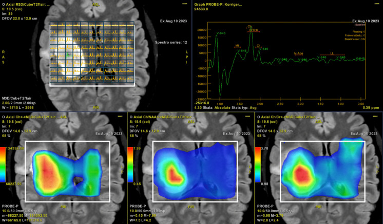
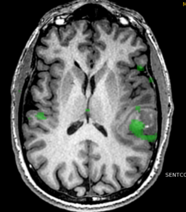
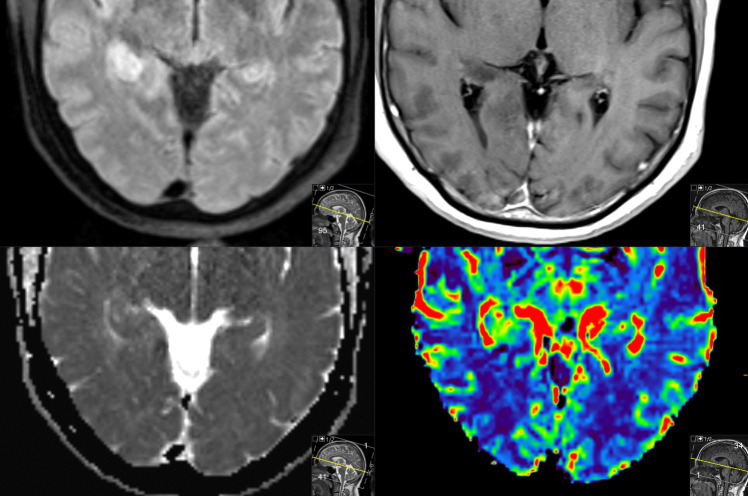
Glial tumors and especially glioblastoma present a major challenge in neuro-oncology due to their infiltrative growth, resistance to therapy, and poor overall survival-despite aggressive treatments such as maximal safe resection and chemoradiotherapy. These tumors typically manifest through neurological symptoms such as seizures, headaches, and signs of increased intracranial pressure, prompting urgent neuroimaging. At initial diagnosis, MRI plays a central role in differentiating true neoplasms from tumor mimics, including inflammatory or infectious conditions. Advanced techniques such as perfusion-weighted imaging (PWI) and diffusion-weighted imaging (DWI) enhance diagnostic specificity and may prevent unnecessary surgical intervention. In the preoperative phase, MRI contributes to surgical planning through the use of functional MRI (fMRI) and diffusion tensor imaging (DTI), enabling localization of eloquent cortex and white matter tracts. These modalities support safer resections by informing trajectory planning and risk assessment. Emerging MR techniques, including magnetic resonance spectroscopy, amide proton transfer imaging, and 2HG quantification, offer further potential in delineating tumor infiltration beyond contrast-enhancing margins. Postoperatively, MRI is important for evaluating residual tumor, detecting surgical complications, and guiding radiotherapy planning. During treatment surveillance, MRI assists in distinguishing true progression from pseudoprogression or radiation necrosis, thereby guiding decisions on additional surgery, changes in systemic therapy, or inclusion into clinical trials. The continued evolution of MRI hardware, software, and image analysis-particularly with the integration of machine learning-will be critical for supporting precision neurosurgical oncology. This review highlights how advanced MRI techniques can inform clinical decision-making at each stage of care in patients with high-grade gliomas.
期刊介绍:
The journal "Acta Neurochirurgica" publishes only original papers useful both to research and clinical work. Papers should deal with clinical neurosurgery - diagnosis and diagnostic techniques, operative surgery and results, postoperative treatment - or with research work in neuroscience if the underlying questions or the results are of neurosurgical interest. Reports on congresses are given in brief accounts. As official organ of the European Association of Neurosurgical Societies the journal publishes all announcements of the E.A.N.S. and reports on the activities of its member societies. Only contributions written in English will be accepted.

 求助内容:
求助内容: 应助结果提醒方式:


