Paul Yousif, Forrest Linch, Prabhakar Rajiah, Jeremy D Collins, Christopher P Favazza, Andrea Ferrero, Michael Jundt, Scott Thompson
{"title":"Photon counting detector CTA for prostate artery embolization pre-procedure planning and intra-procedural guidance.","authors":"Paul Yousif, Forrest Linch, Prabhakar Rajiah, Jeremy D Collins, Christopher P Favazza, Andrea Ferrero, Michael Jundt, Scott Thompson","doi":"10.1186/s42155-025-00567-6","DOIUrl":null,"url":null,"abstract":"<p><strong>Background: </strong>Prostate artery embolization (PAE) requires a careful understanding of pelvic arterial anatomy and identifying prostatic artery variants. Pre-procedure CTA and intra-procedural cone beam CT are traditional means of planning and performing PAE, with the latter providing guidance for embolization. Photon counting detector (PCD) CT enables ultra-high spatial resolution (UHR) whole-body imaging. For PAE, we obtain a single UHR PCD CT arterial phase acquisition, which provides both detailed pre-procedure pelvic arterial anatomic information and a dataset for 2D (angiographic) to 3D (CTA) fusion for intra-procedural guidance during PAE using embolization guidance software in the angiography suite.</p><p><strong>Case presentations: </strong>In six patients who underwent technically successful PAE via a left transradial approach, the pre-procedure diagnostic UHR pelvic PCD prostate CTA delineated bilateral prostatic artery origins and course in all cases, as confirmed with conventional angiograms. Further, registration of the UHR PCD CTA for embolization guidance was successful in all cases, augmenting vessel selection. No complication occurred.</p><p><strong>Conclusion: </strong>UHR PCD CTA is a novel imaging technology that can provide detailed prostate arterial anatomic information for pre-procedure PAE planning. Further, this same UHR PCD CTA dataset can be used for intra-procedural embolization guidance using commercially available embolization guidance software.</p>","PeriodicalId":52351,"journal":{"name":"CVIR Endovascular","volume":"8 1","pages":"55"},"PeriodicalIF":1.5000,"publicationDate":"2025-06-21","publicationTypes":"Journal Article","fieldsOfStudy":null,"isOpenAccess":false,"openAccessPdf":"https://www.ncbi.nlm.nih.gov/pmc/articles/PMC12181139/pdf/","citationCount":"0","resultStr":null,"platform":"Semanticscholar","paperid":null,"PeriodicalName":"CVIR Endovascular","FirstCategoryId":"1085","ListUrlMain":"https://doi.org/10.1186/s42155-025-00567-6","RegionNum":0,"RegionCategory":null,"ArticlePicture":[],"TitleCN":null,"AbstractTextCN":null,"PMCID":null,"EPubDate":"","PubModel":"","JCR":"Q3","JCRName":"CARDIAC & CARDIOVASCULAR SYSTEMS","Score":null,"Total":0}
引用次数: 0
Abstract
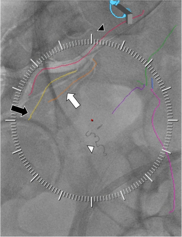
Background: Prostate artery embolization (PAE) requires a careful understanding of pelvic arterial anatomy and identifying prostatic artery variants. Pre-procedure CTA and intra-procedural cone beam CT are traditional means of planning and performing PAE, with the latter providing guidance for embolization. Photon counting detector (PCD) CT enables ultra-high spatial resolution (UHR) whole-body imaging. For PAE, we obtain a single UHR PCD CT arterial phase acquisition, which provides both detailed pre-procedure pelvic arterial anatomic information and a dataset for 2D (angiographic) to 3D (CTA) fusion for intra-procedural guidance during PAE using embolization guidance software in the angiography suite.
Case presentations: In six patients who underwent technically successful PAE via a left transradial approach, the pre-procedure diagnostic UHR pelvic PCD prostate CTA delineated bilateral prostatic artery origins and course in all cases, as confirmed with conventional angiograms. Further, registration of the UHR PCD CTA for embolization guidance was successful in all cases, augmenting vessel selection. No complication occurred.
Conclusion: UHR PCD CTA is a novel imaging technology that can provide detailed prostate arterial anatomic information for pre-procedure PAE planning. Further, this same UHR PCD CTA dataset can be used for intra-procedural embolization guidance using commercially available embolization guidance software.
 求助内容:
求助内容: 应助结果提醒方式:


