Shaun G Hong, Sang Mok Park, Semin Kwon, Haripriya Sakthivel, Jung Woo Leem, Steven R Steinhubl, Pascal Ngiruwonsanga, Jean-Louis N Mangara, Célestin Twizere, Young L Kim
{"title":"Radiomic identification of anemia features in monochromatic conjunctiva photographs in school-age children.","authors":"Shaun G Hong, Sang Mok Park, Semin Kwon, Haripriya Sakthivel, Jung Woo Leem, Steven R Steinhubl, Pascal Ngiruwonsanga, Jean-Louis N Mangara, Célestin Twizere, Young L Kim","doi":"10.1117/1.bios.2.2.022303","DOIUrl":null,"url":null,"abstract":"<p><strong>Significance: </strong>Anemia remains a substantial global health challenge. Delayed detection often leads to various health complications. In school-age children, anemia can impair both cognitive and physical development. Timely detection is particularly critical for this vulnerable population as effective interventions are available even in resource-limited settings.</p><p><strong>Aim: </strong>Most existing methods for assessing conjunctiva paleness or redness in anemia detection rely on colorimetric analyses or spectral imaging, which require sophisticated color processing methods or specialized equipment. We introduce an alternative that takes advantage of purely spatial and textural characteristics of the conjunctiva microvasculature for anemia detection.</p><p><strong>Approach: </strong>Radiomics, an emerging machine learning approach for conventional medical imaging, is applied to conjunctiva photos to analyze morphological alterations in the microvasculature beyond direct visualization. Radiomic analyses are conducted on 12,441 palpebral and 12,375 bulbar conjunctiva photos, captured using three different smartphone models from 565 children aged 5 to 15 years.</p><p><strong>Results: </strong>Spatial and textural features extracted from the palpebral and bulbar conjunctivae are significantly associated with anemia status in school-age children, demonstrating their potential as biomarkers of anemia.</p><p><strong>Conclusions: </strong>Instead of relying on color-based or spectral analyses of pallor in the conjunctiva, the proposed framework lays the groundwork for simplifying the hardware and algorithmic requirements of point-of-care, noninvasive anemia screening in sub-Saharan Africa and other resource-limited settings.</p>","PeriodicalId":519981,"journal":{"name":"Biophotonics discovery","volume":"2 2","pages":""},"PeriodicalIF":0.0000,"publicationDate":"2025-04-01","publicationTypes":"Journal Article","fieldsOfStudy":null,"isOpenAccess":false,"openAccessPdf":"https://www.ncbi.nlm.nih.gov/pmc/articles/PMC12176424/pdf/","citationCount":"0","resultStr":null,"platform":"Semanticscholar","paperid":null,"PeriodicalName":"Biophotonics discovery","FirstCategoryId":"1085","ListUrlMain":"https://doi.org/10.1117/1.bios.2.2.022303","RegionNum":0,"RegionCategory":null,"ArticlePicture":[],"TitleCN":null,"AbstractTextCN":null,"PMCID":null,"EPubDate":"2025/4/15 0:00:00","PubModel":"Epub","JCR":"","JCRName":"","Score":null,"Total":0}
引用次数: 0
Abstract
Significance: Anemia remains a substantial global health challenge. Delayed detection often leads to various health complications. In school-age children, anemia can impair both cognitive and physical development. Timely detection is particularly critical for this vulnerable population as effective interventions are available even in resource-limited settings.
Aim: Most existing methods for assessing conjunctiva paleness or redness in anemia detection rely on colorimetric analyses or spectral imaging, which require sophisticated color processing methods or specialized equipment. We introduce an alternative that takes advantage of purely spatial and textural characteristics of the conjunctiva microvasculature for anemia detection.
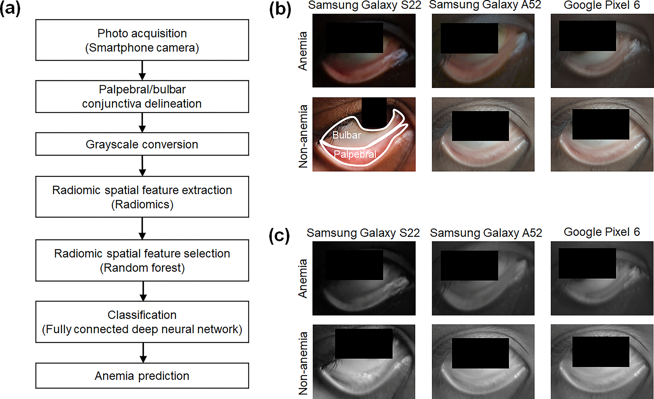
Approach: Radiomics, an emerging machine learning approach for conventional medical imaging, is applied to conjunctiva photos to analyze morphological alterations in the microvasculature beyond direct visualization. Radiomic analyses are conducted on 12,441 palpebral and 12,375 bulbar conjunctiva photos, captured using three different smartphone models from 565 children aged 5 to 15 years.
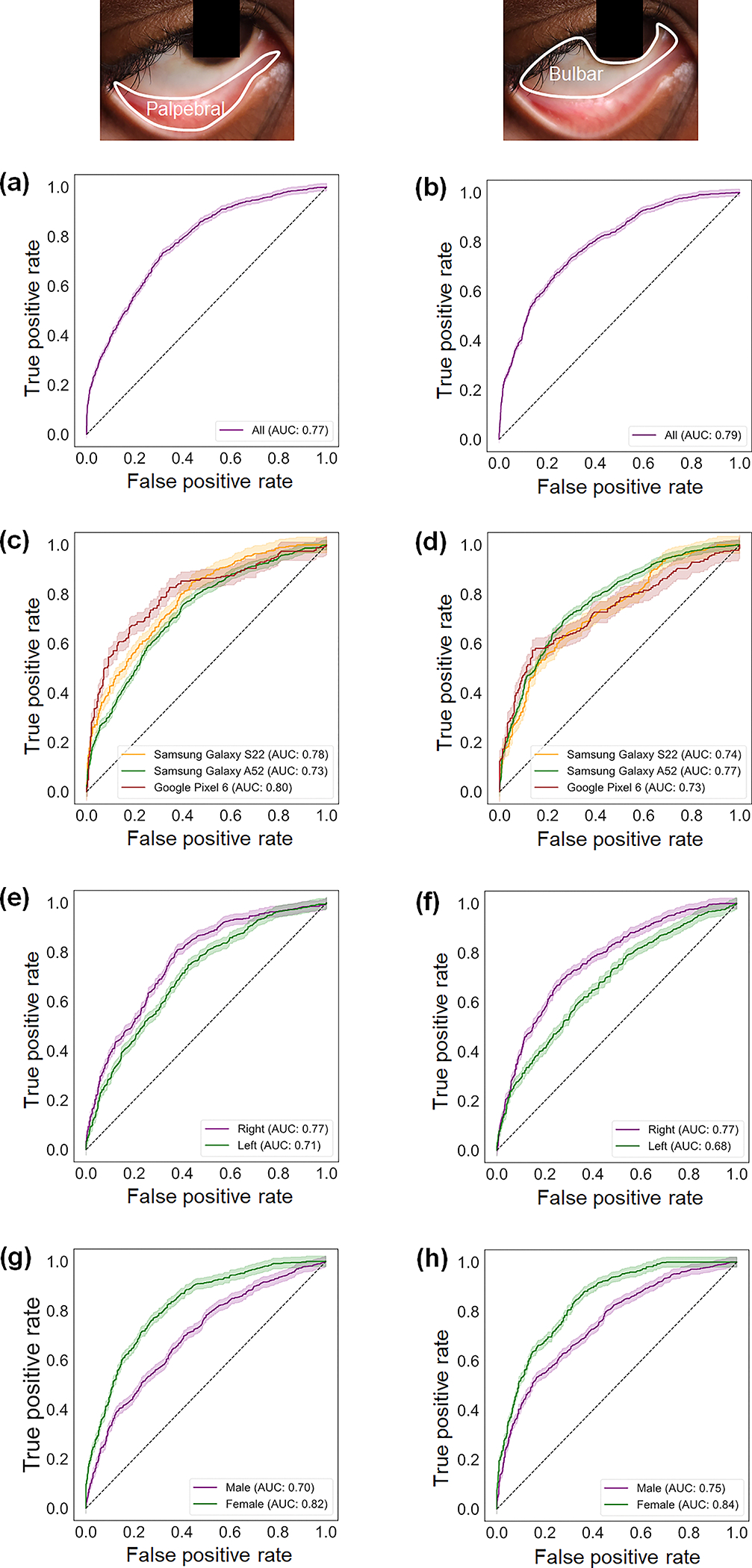
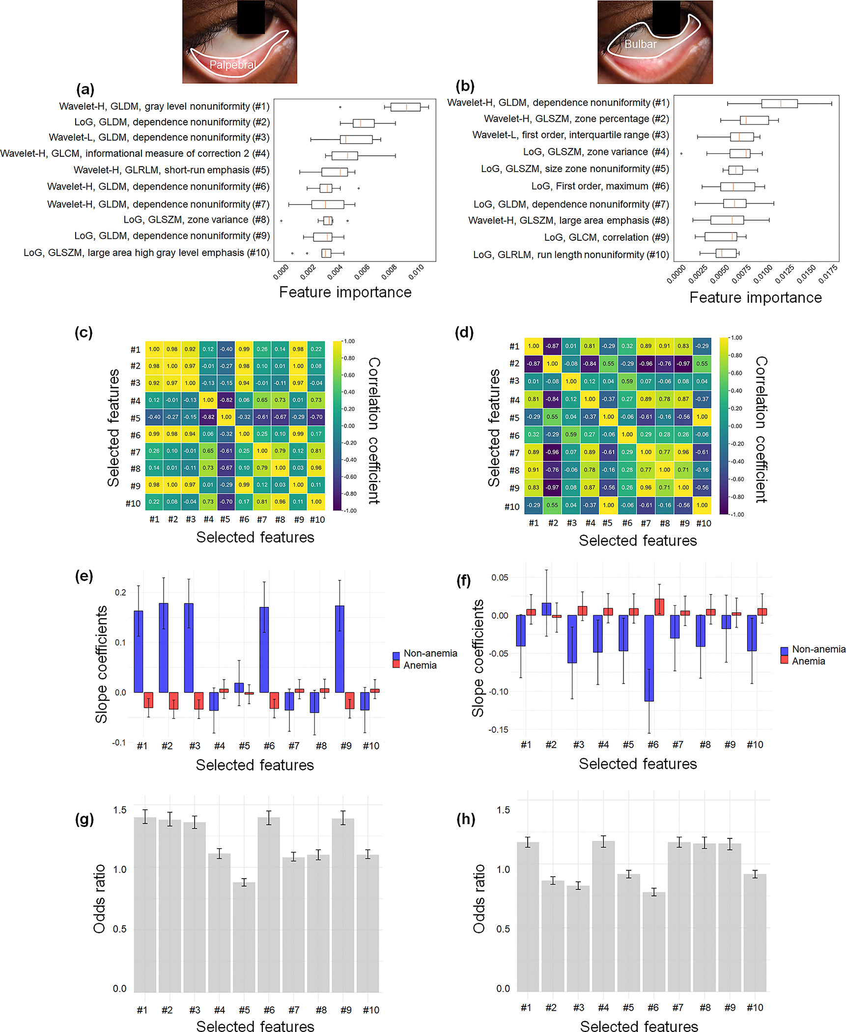
Results: Spatial and textural features extracted from the palpebral and bulbar conjunctivae are significantly associated with anemia status in school-age children, demonstrating their potential as biomarkers of anemia.
Conclusions: Instead of relying on color-based or spectral analyses of pallor in the conjunctiva, the proposed framework lays the groundwork for simplifying the hardware and algorithmic requirements of point-of-care, noninvasive anemia screening in sub-Saharan Africa and other resource-limited settings.

 求助内容:
求助内容: 应助结果提醒方式:


