Yucens Mehmet, Aydemir Ahmet Nadir, Funda Fatma Bolukbası Hatip, Zeynep Mine Altunay, Gülcin Abban Mete, Mehmet Bilgen, Fahir Demirkan
{"title":"Effects of traumatic brain injury on vascular response and fracture healing: an experimental study in a rat model.","authors":"Yucens Mehmet, Aydemir Ahmet Nadir, Funda Fatma Bolukbası Hatip, Zeynep Mine Altunay, Gülcin Abban Mete, Mehmet Bilgen, Fahir Demirkan","doi":"10.5152/j.aott.2025.23104","DOIUrl":null,"url":null,"abstract":"<p><p>Objective: This study aimed to investigate the effects of traumatic brain injury (TBI) on vascular response and fracture healing during recovery. Methods: In this experimental animal study, a total of 63 male Wistar albino rats (200-250 g) were randomly assigned to 3 groups: TBI with tibia fracture (TBI+Fx, n=21), tibia fracture only (Fx only, n=21), and a control group (n=21). Traumatic brain injury was induced in the motor cortex using a controlled impact device, followed by the tibia fracture. The severity of TBI was assessed using rotarod tests. Blood samples were collected on days 1, 7, and 21 post-fracture, while brain and tibia samples were taken on day 21 following decapitation. Levels of antidiuretic hormone (ADH) and angiotensin 1-7 (Ang 1-7) were quantified using Enzyme-linked immunosorbent assays (ELISA). Fracture healing was assessed through micro-CT and histopathological analysis. Aortic segments were evaluated for contractile response and relaxation in isolated organ baths. Results: Micro-CT analysis revealed significantly greater bone volume (BV) (P=.02) and trabecular number (P=.038) in the TBI+Fx group. Histopathological healing scores were also significantly higher in the TBI+Fx group compared to the Fx only group (P=.019). Potassium chloride (KCl) induced contractile responses were greater in the Fx only group than in the TBI+Fx group (P < .05). Acetylcholine (ACh) induced relaxation was diminished in both Fx and TBI+Fx groups compared to controls (P < .01), whereas sodium nitroprusside (SNP)-induced relaxation was significantly greater in the TBI+Fx group than in the Fx only and control groups (P < .05). On day 21, arginine vasopressin (AVP) levels were significantly higher in the Fx only group compared to the TBI+Fx group (P=.034), with no significant differences observed on days 1 and 7. Plasma Ang 1-7 levels were significantly elevated in the Fx only group on day 21 compared to the TBI+Fx group (P < .05). Conclusion: Traumatic brain injury was associated with accelerated fracture healing, as evidenced by increased BV, trabecular thickness, and histopathological healing scores. Additionally, TBI appeared to modulate vascular function, possibly via mechanisms involving nitric oxide and calcium signaling. These findings suggest that neuroendocrine changes following TBI may enhance fracture healing, offering potential clinical insights for managing polytrauma patients. Level of Evidence: N/A.</p>","PeriodicalId":93854,"journal":{"name":"Acta orthopaedica et traumatologica turcica","volume":"59 3","pages":"133-140"},"PeriodicalIF":1.0000,"publicationDate":"2025-05-28","publicationTypes":"Journal Article","fieldsOfStudy":null,"isOpenAccess":false,"openAccessPdf":"https://www.ncbi.nlm.nih.gov/pmc/articles/PMC12147349/pdf/","citationCount":"0","resultStr":null,"platform":"Semanticscholar","paperid":null,"PeriodicalName":"Acta orthopaedica et traumatologica turcica","FirstCategoryId":"1085","ListUrlMain":"https://doi.org/10.5152/j.aott.2025.23104","RegionNum":0,"RegionCategory":null,"ArticlePicture":[],"TitleCN":null,"AbstractTextCN":null,"PMCID":null,"EPubDate":"","PubModel":"","JCR":"","JCRName":"","Score":null,"Total":0}
引用次数: 0
Abstract
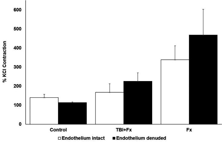
Objective: This study aimed to investigate the effects of traumatic brain injury (TBI) on vascular response and fracture healing during recovery. Methods: In this experimental animal study, a total of 63 male Wistar albino rats (200-250 g) were randomly assigned to 3 groups: TBI with tibia fracture (TBI+Fx, n=21), tibia fracture only (Fx only, n=21), and a control group (n=21). Traumatic brain injury was induced in the motor cortex using a controlled impact device, followed by the tibia fracture. The severity of TBI was assessed using rotarod tests. Blood samples were collected on days 1, 7, and 21 post-fracture, while brain and tibia samples were taken on day 21 following decapitation. Levels of antidiuretic hormone (ADH) and angiotensin 1-7 (Ang 1-7) were quantified using Enzyme-linked immunosorbent assays (ELISA). Fracture healing was assessed through micro-CT and histopathological analysis. Aortic segments were evaluated for contractile response and relaxation in isolated organ baths. Results: Micro-CT analysis revealed significantly greater bone volume (BV) (P=.02) and trabecular number (P=.038) in the TBI+Fx group. Histopathological healing scores were also significantly higher in the TBI+Fx group compared to the Fx only group (P=.019). Potassium chloride (KCl) induced contractile responses were greater in the Fx only group than in the TBI+Fx group (P < .05). Acetylcholine (ACh) induced relaxation was diminished in both Fx and TBI+Fx groups compared to controls (P < .01), whereas sodium nitroprusside (SNP)-induced relaxation was significantly greater in the TBI+Fx group than in the Fx only and control groups (P < .05). On day 21, arginine vasopressin (AVP) levels were significantly higher in the Fx only group compared to the TBI+Fx group (P=.034), with no significant differences observed on days 1 and 7. Plasma Ang 1-7 levels were significantly elevated in the Fx only group on day 21 compared to the TBI+Fx group (P < .05). Conclusion: Traumatic brain injury was associated with accelerated fracture healing, as evidenced by increased BV, trabecular thickness, and histopathological healing scores. Additionally, TBI appeared to modulate vascular function, possibly via mechanisms involving nitric oxide and calcium signaling. These findings suggest that neuroendocrine changes following TBI may enhance fracture healing, offering potential clinical insights for managing polytrauma patients. Level of Evidence: N/A.


 求助内容:
求助内容: 应助结果提醒方式:


