Structural Connectivity of the Basal Ganglia from Patient-Individual Tractography Is Key for Understanding the Effects of Deep Brain Stimulation in Parkinson's Disease.
Ricardo Loução, Martin Kocher, Gregor Alexander Brandt, Jan Niklas Petry-Schmelzer, Michael Barbe, Haidar Dafsari, Josef Mana, Robert Jech, Jochen Wirths, Veerle Visser-Vandewalle, Pablo Andrade, Halim Baqapuri, Michael Luehrs, David E J Linden, Brendan Santyr, Andres Lozano, Alessandro Bongioanni, Bechir Jarraya, Tolga Cukur
{"title":"Structural Connectivity of the Basal Ganglia from Patient-Individual Tractography Is Key for Understanding the Effects of Deep Brain Stimulation in Parkinson's Disease.","authors":"Ricardo Loução, Martin Kocher, Gregor Alexander Brandt, Jan Niklas Petry-Schmelzer, Michael Barbe, Haidar Dafsari, Josef Mana, Robert Jech, Jochen Wirths, Veerle Visser-Vandewalle, Pablo Andrade, Halim Baqapuri, Michael Luehrs, David E J Linden, Brendan Santyr, Andres Lozano, Alessandro Bongioanni, Bechir Jarraya, Tolga Cukur","doi":"10.1159/000546716","DOIUrl":null,"url":null,"abstract":"<p><strong>Introduction: </strong>In Parkinson's disease (PD) patients, modulation of the fibre tracts of the cortico-basal ganglia-thalamo-cortical loop is the presumed mechanism of action of deep brain stimulation (DBS) of the subthalamic nucleus (STN). Therefore, we explored patient-individual cortical structural connectivity of the volume of tissue activated (VTA), as well as DBS-induced modulation of fibre tracts connecting the STN with cortical and subcortical nodes, and their correlation with therapeutic effects.</p><p><strong>Methods: </strong>A retrospective cohort of n = 69 PD patients treated with bilateral DBS of the STN was analysed. Clinical response was assessed from the DBS-induced change in the UPDRS-III motor scores (total and symptom-specific sub-scores) under regular medication after a median follow-up of 9.0 (range 2.6-20.2) months. Tractography based on patient-individual diffusion-weighted MRI was employed in two ways. Whole-brain tractography was used to identify the cortical connections of fibres passing the VTAs, and reconstruction of specific white matter pathways of the motor loop connecting the STN with the basal ganglia and cortex was used to identify the proportion of fibres within these pathways which was modulated by STN-DBS. This proportion of pathway modulation was used in a correlative analysis with clinical outcomes.</p><p><strong>Results: </strong>Fibres traversing the VTAs were primarily connected to the supplementary motor area (SMA) and to a lesser degree to the premotor cortex. Within the pathways connecting the STN with the cortical and subcortical nodes, on average 30-40% (range 10-80%) of the fibres were modulated by STN-DBS. This proportion correlated significantly with the percentage change in UPDRS motor score for fibres connecting the STN with the SMA (ρ = 0.28), pre-SMA (ρ = 0.26), ventral and dorsal premotor cortices (ρ = 0.26 and ρ = 0.29, respectively), and the globus pallidus externus (ρ = 0.26) and internus (ρ = 0.29). Also, good clinical responses for both tremor and rigidity were associated with a significantly (p < 0.05) higher proportion of modulated fibres for the same cortico- and sub-cortico-STN connections.</p><p><strong>Conclusion: </strong>Patient-individual tractography reveals that, in PD, most of the cortical fibres traversing the VTA are connected to the SMA. In addition, clinical efficacy is related to the proportion of DBS-affected fibres connecting the STN with nodes of both the hyperdirect (cortex-STN) and the indirect pathways (STN-basal ganglia). As such, patient-specific tractography, in particular in the basal ganglia, could be used in a clinical context as a tool to guide therapy.</p>","PeriodicalId":22078,"journal":{"name":"Stereotactic and Functional Neurosurgery","volume":" ","pages":"279-294"},"PeriodicalIF":2.4000,"publicationDate":"2025-01-01","publicationTypes":"Journal Article","fieldsOfStudy":null,"isOpenAccess":false,"openAccessPdf":"https://www.ncbi.nlm.nih.gov/pmc/articles/PMC12453574/pdf/","citationCount":"0","resultStr":null,"platform":"Semanticscholar","paperid":null,"PeriodicalName":"Stereotactic and Functional Neurosurgery","FirstCategoryId":"3","ListUrlMain":"https://doi.org/10.1159/000546716","RegionNum":4,"RegionCategory":"医学","ArticlePicture":[],"TitleCN":null,"AbstractTextCN":null,"PMCID":null,"EPubDate":"2025/6/18 0:00:00","PubModel":"Epub","JCR":"Q3","JCRName":"NEUROIMAGING","Score":null,"Total":0}
引用次数: 0
Abstract
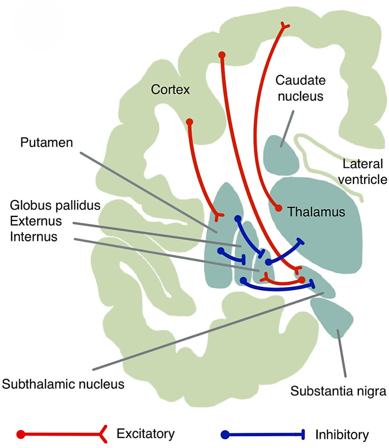
Introduction: In Parkinson's disease (PD) patients, modulation of the fibre tracts of the cortico-basal ganglia-thalamo-cortical loop is the presumed mechanism of action of deep brain stimulation (DBS) of the subthalamic nucleus (STN). Therefore, we explored patient-individual cortical structural connectivity of the volume of tissue activated (VTA), as well as DBS-induced modulation of fibre tracts connecting the STN with cortical and subcortical nodes, and their correlation with therapeutic effects.
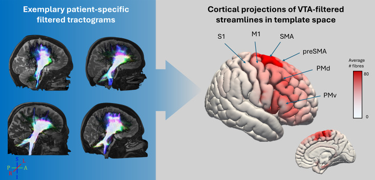
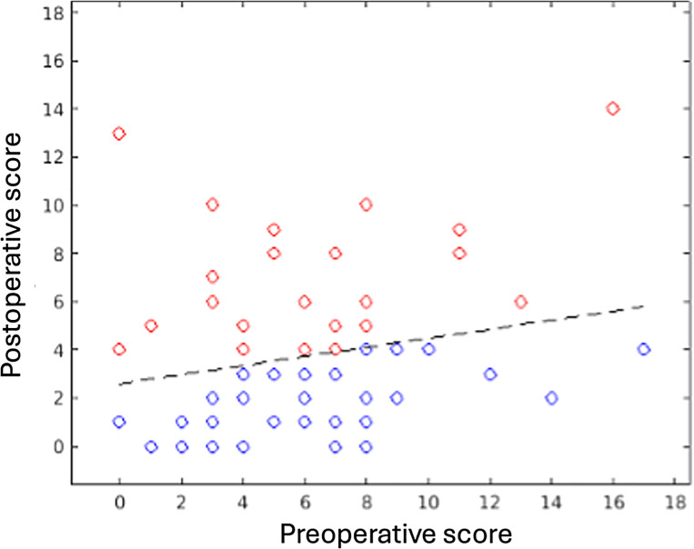
Methods: A retrospective cohort of n = 69 PD patients treated with bilateral DBS of the STN was analysed. Clinical response was assessed from the DBS-induced change in the UPDRS-III motor scores (total and symptom-specific sub-scores) under regular medication after a median follow-up of 9.0 (range 2.6-20.2) months. Tractography based on patient-individual diffusion-weighted MRI was employed in two ways. Whole-brain tractography was used to identify the cortical connections of fibres passing the VTAs, and reconstruction of specific white matter pathways of the motor loop connecting the STN with the basal ganglia and cortex was used to identify the proportion of fibres within these pathways which was modulated by STN-DBS. This proportion of pathway modulation was used in a correlative analysis with clinical outcomes.
Results: Fibres traversing the VTAs were primarily connected to the supplementary motor area (SMA) and to a lesser degree to the premotor cortex. Within the pathways connecting the STN with the cortical and subcortical nodes, on average 30-40% (range 10-80%) of the fibres were modulated by STN-DBS. This proportion correlated significantly with the percentage change in UPDRS motor score for fibres connecting the STN with the SMA (ρ = 0.28), pre-SMA (ρ = 0.26), ventral and dorsal premotor cortices (ρ = 0.26 and ρ = 0.29, respectively), and the globus pallidus externus (ρ = 0.26) and internus (ρ = 0.29). Also, good clinical responses for both tremor and rigidity were associated with a significantly (p < 0.05) higher proportion of modulated fibres for the same cortico- and sub-cortico-STN connections.
Conclusion: Patient-individual tractography reveals that, in PD, most of the cortical fibres traversing the VTA are connected to the SMA. In addition, clinical efficacy is related to the proportion of DBS-affected fibres connecting the STN with nodes of both the hyperdirect (cortex-STN) and the indirect pathways (STN-basal ganglia). As such, patient-specific tractography, in particular in the basal ganglia, could be used in a clinical context as a tool to guide therapy.
期刊介绍:
''Stereotactic and Functional Neurosurgery'' provides a single source for the reader to keep abreast of developments in the most rapidly advancing subspecialty within neurosurgery. Technological advances in computer-assisted surgery, robotics, imaging and neurophysiology are being applied to clinical problems with ever-increasing rapidity in stereotaxis more than any other field, providing opportunities for new approaches to surgical and radiotherapeutic management of diseases of the brain, spinal cord, and spine. Issues feature advances in the use of deep-brain stimulation, imaging-guided techniques in stereotactic biopsy and craniotomy, stereotactic radiosurgery, and stereotactically implanted and guided radiotherapeutics and biologicals in the treatment of functional and movement disorders, brain tumors, and other diseases of the brain. Background information from basic science laboratories related to such clinical advances provides the reader with an overall perspective of this field. Proceedings and abstracts from many of the key international meetings furnish an overview of this specialty available nowhere else. ''Stereotactic and Functional Neurosurgery'' meets the information needs of both investigators and clinicians in this rapidly advancing field.
 求助内容:
求助内容: 应助结果提醒方式:


