Jia Li, Cong Wei, Tao Ying, Yan Liu, Ronghui Wang, Maoyao Li, Chao Feng, Di Sun, Yuanyi Zheng
{"title":"Differentiation of benign and malignant breast lesions by ultrasound localization microscopy.","authors":"Jia Li, Cong Wei, Tao Ying, Yan Liu, Ronghui Wang, Maoyao Li, Chao Feng, Di Sun, Yuanyi Zheng","doi":"10.1186/s13244-025-02013-6","DOIUrl":null,"url":null,"abstract":"<p><strong>Objective: </strong>We investigated the role of ultrasound localization microscopy (ULM) qualitative and quantitative parameters in distinguishing benign from malignant breast lesions.</p><p><strong>Methods: </strong>The ULM qualitative and quantitative parameters of breast lesions were recorded. A receiver operating characteristic (ROC) curve was applied to assess the diagnostic performance of ULM. Intra- and inter-operator reliabilities of quantitative parameters were assessed.</p><p><strong>Results: </strong>Thirty-one breast lesions were verified by pathologic results, 14 of which were benign and 17 were malignant. Benign lesions were associated with dot-like, line-like, or branch-like patterns (93% vs 6%), whereas malignant lesions were associated with chaotic patterns (94% vs 7%) (p < 0.001). The microvasculature morphology had an area under the curve (AUC) of 0.935, a sensitivity of 94.1%, and a specificity of 92.9%. The microvasculature density, mean diameter, largest diameter, and max tortuosity of malignant lesions were significantly greater than those of benign lesions (p < 0.05, p < 0.001, p < 0.001, p < 0.05). The microvasculature mean flow velocity of benign lesions was significantly greater than that of malignant lesions (p < 0.05). For the quantitative parameters, the AUC was highest for the microvasculature's largest diameter (0.962), with a sensitivity of 88.2% and a specificity of 92.9%. The intra- and inter-operator reliabilities of quantitative parameters were excellent (ICC greater than 0.90).</p><p><strong>Conclusions: </strong>ULM is useful for distinguishing benign from malignant breast lesions. ULM can offer a new diagnostic method for breast lesions, which deserves further research.</p><p><strong>Critical relevance statement: </strong>This study suggests that ULM is a new technology with super-resolution that is helpful for distinguishing benign from malignant breast lesions.</p><p><strong>Trial registration: </strong>ChiCTR, ChiCTR2100048361. Registered 6 July 2021, https://www.chictr.org.cn/ .</p><p><strong>Key points: </strong>ULM is an emerging technology that can detect highly detailed networks of microvasculature. Microvasculature morphology based on ULM can be a good indicator for the differential diagnosis of breast lesions. Among quantitative parameters extracted from ULM, microvasculature largest diameter was the best for the classification of breast lesions.</p>","PeriodicalId":13639,"journal":{"name":"Insights into Imaging","volume":"16 1","pages":"128"},"PeriodicalIF":4.5000,"publicationDate":"2025-06-18","publicationTypes":"Journal Article","fieldsOfStudy":null,"isOpenAccess":false,"openAccessPdf":"https://www.ncbi.nlm.nih.gov/pmc/articles/PMC12176706/pdf/","citationCount":"0","resultStr":null,"platform":"Semanticscholar","paperid":null,"PeriodicalName":"Insights into Imaging","FirstCategoryId":"3","ListUrlMain":"https://doi.org/10.1186/s13244-025-02013-6","RegionNum":2,"RegionCategory":"医学","ArticlePicture":[],"TitleCN":null,"AbstractTextCN":null,"PMCID":null,"EPubDate":"","PubModel":"","JCR":"Q1","JCRName":"RADIOLOGY, NUCLEAR MEDICINE & MEDICAL IMAGING","Score":null,"Total":0}
引用次数: 0
Abstract
Objective: We investigated the role of ultrasound localization microscopy (ULM) qualitative and quantitative parameters in distinguishing benign from malignant breast lesions.
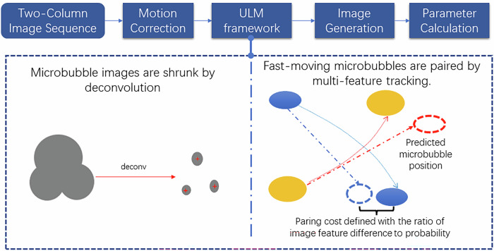
Methods: The ULM qualitative and quantitative parameters of breast lesions were recorded. A receiver operating characteristic (ROC) curve was applied to assess the diagnostic performance of ULM. Intra- and inter-operator reliabilities of quantitative parameters were assessed.
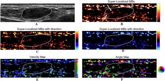
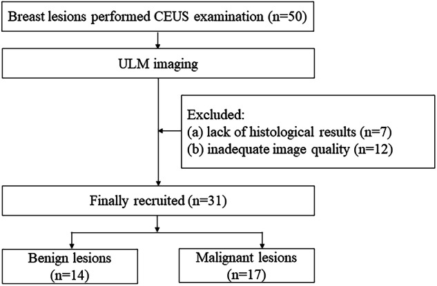
Results: Thirty-one breast lesions were verified by pathologic results, 14 of which were benign and 17 were malignant. Benign lesions were associated with dot-like, line-like, or branch-like patterns (93% vs 6%), whereas malignant lesions were associated with chaotic patterns (94% vs 7%) (p < 0.001). The microvasculature morphology had an area under the curve (AUC) of 0.935, a sensitivity of 94.1%, and a specificity of 92.9%. The microvasculature density, mean diameter, largest diameter, and max tortuosity of malignant lesions were significantly greater than those of benign lesions (p < 0.05, p < 0.001, p < 0.001, p < 0.05). The microvasculature mean flow velocity of benign lesions was significantly greater than that of malignant lesions (p < 0.05). For the quantitative parameters, the AUC was highest for the microvasculature's largest diameter (0.962), with a sensitivity of 88.2% and a specificity of 92.9%. The intra- and inter-operator reliabilities of quantitative parameters were excellent (ICC greater than 0.90).
Conclusions: ULM is useful for distinguishing benign from malignant breast lesions. ULM can offer a new diagnostic method for breast lesions, which deserves further research.
Critical relevance statement: This study suggests that ULM is a new technology with super-resolution that is helpful for distinguishing benign from malignant breast lesions.
Trial registration: ChiCTR, ChiCTR2100048361. Registered 6 July 2021, https://www.chictr.org.cn/ .
Key points: ULM is an emerging technology that can detect highly detailed networks of microvasculature. Microvasculature morphology based on ULM can be a good indicator for the differential diagnosis of breast lesions. Among quantitative parameters extracted from ULM, microvasculature largest diameter was the best for the classification of breast lesions.
期刊介绍:
Insights into Imaging (I³) is a peer-reviewed open access journal published under the brand SpringerOpen. All content published in the journal is freely available online to anyone, anywhere!
I³ continuously updates scientific knowledge and progress in best-practice standards in radiology through the publication of original articles and state-of-the-art reviews and opinions, along with recommendations and statements from the leading radiological societies in Europe.
Founded by the European Society of Radiology (ESR), I³ creates a platform for educational material, guidelines and recommendations, and a forum for topics of controversy.
A balanced combination of review articles, original papers, short communications from European radiological congresses and information on society matters makes I³ an indispensable source for current information in this field.
I³ is owned by the ESR, however authors retain copyright to their article according to the Creative Commons Attribution License (see Copyright and License Agreement). All articles can be read, redistributed and reused for free, as long as the author of the original work is cited properly.
The open access fees (article-processing charges) for this journal are kindly sponsored by ESR for all Members.
The journal went open access in 2012, which means that all articles published since then are freely available online.

 求助内容:
求助内容: 应助结果提醒方式:


