Dong Chen, Jian Hong, Wenting Li, Qiuju Sheng, Olivia Mezzetti, Min Xu, Shadi Salloum, Raymond T Chung, Wenyu Lin
{"title":"HBV promotes epithelial-mesenchymal transition in HCC and liver fibrosis through JNK-mediated autophagy.","authors":"Dong Chen, Jian Hong, Wenting Li, Qiuju Sheng, Olivia Mezzetti, Min Xu, Shadi Salloum, Raymond T Chung, Wenyu Lin","doi":"10.1097/HC9.0000000000000730","DOIUrl":null,"url":null,"abstract":"<p><strong>Background: </strong>HBV, which contributes to liver fibrosis and HCC, is associated with autophagy and epithelial-mesenchymal transition (EMT). However, the relationship between HBV infection, autophagy, and EMT remains unknown.</p><p><strong>Methods: </strong>In this study, we used HBV cell lines (HepAD38, Huh7.5.1-NTCP) to evaluate this relationship.</p><p><strong>Results: </strong>HBV replication or infection promoted autophagy, EMT, and liver fibrogenesis. HBV-induced EMT/fibrogenesis was dependent on autophagy. HBV X and HBV core (C) were each involved during this process. TGF-β1 partially participated in HBV-induced autophagy/EMT/fibrosis. c-Jun N-terminal kinase (JNK) expression was increased in HepAD38 cells, HBV-X (HBV-C)-transfected Huh7.5.1 cells, and HBV-infected Huh7.5.1-NTCP cells. JNK silencing attenuated HBV-induced autophagy and EMT/fibrosis signaling as well. Moreover, overexpression of HBV-X(C) and augmentation of autophagy were shown to promote pJNK expression. Finally, we confirmed an HBV-JNK-autophagy-EMT/fibrosis signaling pathway in a primary human hepatocyte model.</p><p><strong>Conclusions: </strong>In conclusion, HBV induces autophagy through JNK, thus triggering downstream EMT and fibrogenesis signaling pathways. These findings suggest that JNKs could be potential targets for the treatment of HBV-related liver diseases.</p>","PeriodicalId":12978,"journal":{"name":"Hepatology Communications","volume":"9 7","pages":""},"PeriodicalIF":5.6000,"publicationDate":"2025-06-19","publicationTypes":"Journal Article","fieldsOfStudy":null,"isOpenAccess":false,"openAccessPdf":"https://www.ncbi.nlm.nih.gov/pmc/articles/PMC12180830/pdf/","citationCount":"0","resultStr":null,"platform":"Semanticscholar","paperid":null,"PeriodicalName":"Hepatology Communications","FirstCategoryId":"3","ListUrlMain":"https://doi.org/10.1097/HC9.0000000000000730","RegionNum":2,"RegionCategory":"医学","ArticlePicture":[],"TitleCN":null,"AbstractTextCN":null,"PMCID":null,"EPubDate":"2025/7/1 0:00:00","PubModel":"eCollection","JCR":"Q1","JCRName":"GASTROENTEROLOGY & HEPATOLOGY","Score":null,"Total":0}
引用次数: 0
Abstract
Background: HBV, which contributes to liver fibrosis and HCC, is associated with autophagy and epithelial-mesenchymal transition (EMT). However, the relationship between HBV infection, autophagy, and EMT remains unknown.
Methods: In this study, we used HBV cell lines (HepAD38, Huh7.5.1-NTCP) to evaluate this relationship.
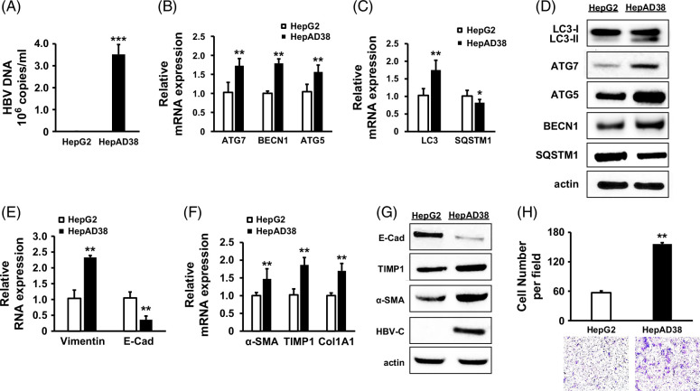
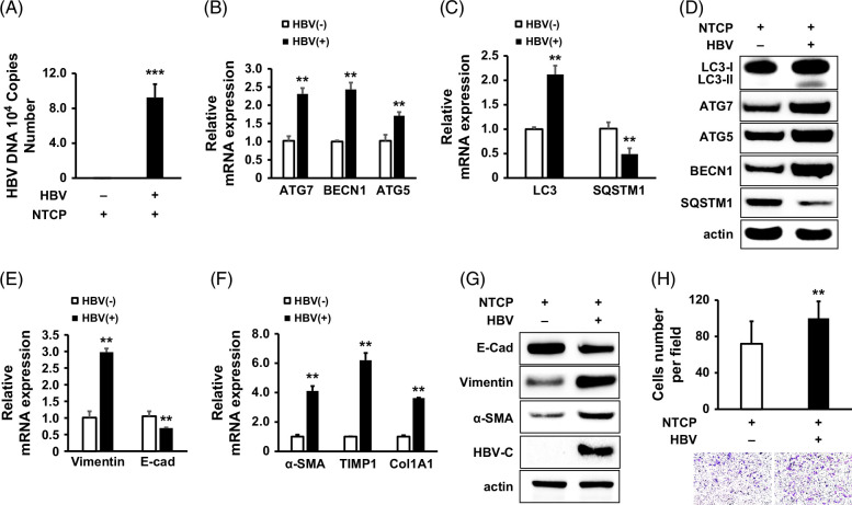
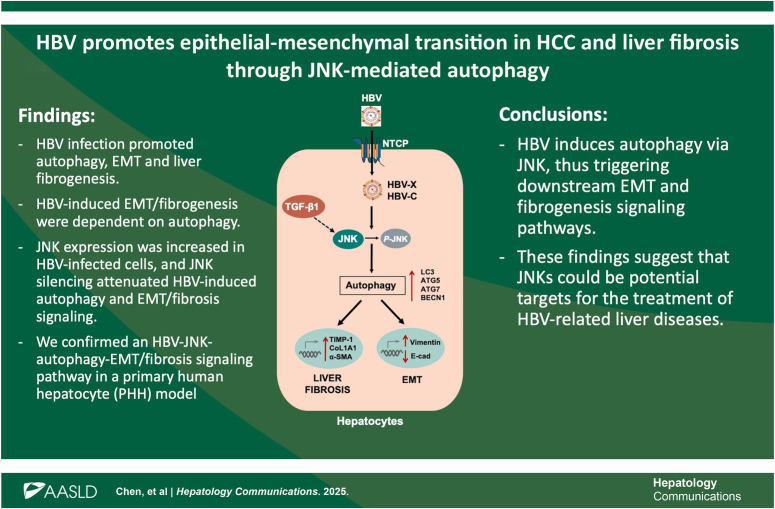
Results: HBV replication or infection promoted autophagy, EMT, and liver fibrogenesis. HBV-induced EMT/fibrogenesis was dependent on autophagy. HBV X and HBV core (C) were each involved during this process. TGF-β1 partially participated in HBV-induced autophagy/EMT/fibrosis. c-Jun N-terminal kinase (JNK) expression was increased in HepAD38 cells, HBV-X (HBV-C)-transfected Huh7.5.1 cells, and HBV-infected Huh7.5.1-NTCP cells. JNK silencing attenuated HBV-induced autophagy and EMT/fibrosis signaling as well. Moreover, overexpression of HBV-X(C) and augmentation of autophagy were shown to promote pJNK expression. Finally, we confirmed an HBV-JNK-autophagy-EMT/fibrosis signaling pathway in a primary human hepatocyte model.
Conclusions: In conclusion, HBV induces autophagy through JNK, thus triggering downstream EMT and fibrogenesis signaling pathways. These findings suggest that JNKs could be potential targets for the treatment of HBV-related liver diseases.
期刊介绍:
Hepatology Communications is a peer-reviewed, online-only, open access journal for fast dissemination of high quality basic, translational, and clinical research in hepatology. Hepatology Communications maintains high standard and rigorous peer review. Because of its open access nature, authors retain the copyright to their works, all articles are immediately available and free to read and share, and it is fully compliant with funder and institutional mandates. The journal is committed to fast publication and author satisfaction.
 求助内容:
求助内容: 应助结果提醒方式:


