Karla Kerlikowske, Linn Abraham, Brian L Sprague, Olivia Sattayapiwat, Sarah J Nyante, Jeffrey A Tice, Diana L Miglioretti
{"title":"Mammographic calcifications association with risk of advanced breast cancer.","authors":"Karla Kerlikowske, Linn Abraham, Brian L Sprague, Olivia Sattayapiwat, Sarah J Nyante, Jeffrey A Tice, Diana L Miglioretti","doi":"10.1007/s10549-025-07753-z","DOIUrl":null,"url":null,"abstract":"<p><strong>Purpose: </strong>Mammographic calcifications on mammograms with a negative/benign assessment are associated with increased breast cancer risk. Associations with advanced breast cancer risk are unknown. We evaluated whether calcifications recorded on mammography reports are associated with advanced invasive breast cancer risk.</p><p><strong>Methods: </strong>We included 3,710,313 screening mammograms with a negative/benign final assessment performed on 991,991 women aged 40-74 in the Breast Cancer Surveillance Consortium associated with 7229 advanced cancers. We calculated cumulative 5-year advanced (prognostic pathologic stage ≥II) breast cancer risk and hazards ratios (HR) adjusted for clinical risk factors according to presence or absence of calcifications by menopausal status, dense (heterogeneously or extremely dense) vs. non-dense (almost entirely fatty or scattered fibroglandular density) breasts, body mass index (BMI) < 25 kg/m<sup>2</sup> vs. ≥ 25 kg/m<sup>2</sup>.</p><p><strong>Results: </strong>Prevalence of calcifications was 6.1% among women who developed advanced breast cancer vs. 3.6% among others. Overall associations of advanced cancer with calcifications were similar for premenopausal (HR = 1.4; 95% CI 1.1-1.9) and postmenopausal (HR = 1.5; 95% CI 1.2-1.7) women. Compared to postmenopausal women with non-dense breasts and BMI < 25 kg/m<sup>2</sup> without calcifications [cumulative 5-year advanced cancer incidence = 1.6 (95% CI 1.3-2.0) per 1000 women], postmenopausal women with dense breasts, BMI ≥ 25 kg/m<sup>2</sup>, and calcifications had 5.5-fold (95% CI 3.9-7.7) higher advanced cancer risk [cumulative 5-year advanced cancer incidence = 10.2; (95% CI 7.0-13.3) per 1000 women]. Results were similar for premenopausal women.</p><p><strong>Conclusion: </strong>Mammographic calcifications increase advanced cancer risk beyond having dense breasts and being overweight/obese. Future research should investigate strength of associations by type of calcification and incorporation of calcifications into advanced cancer risk models for improvement in model accuracy.</p>","PeriodicalId":9133,"journal":{"name":"Breast Cancer Research and Treatment","volume":" ","pages":"555-567"},"PeriodicalIF":3.0000,"publicationDate":"2025-08-01","publicationTypes":"Journal Article","fieldsOfStudy":null,"isOpenAccess":false,"openAccessPdf":"https://www.ncbi.nlm.nih.gov/pmc/articles/PMC12209027/pdf/","citationCount":"0","resultStr":null,"platform":"Semanticscholar","paperid":null,"PeriodicalName":"Breast Cancer Research and Treatment","FirstCategoryId":"3","ListUrlMain":"https://doi.org/10.1007/s10549-025-07753-z","RegionNum":3,"RegionCategory":"医学","ArticlePicture":[],"TitleCN":null,"AbstractTextCN":null,"PMCID":null,"EPubDate":"2025/6/17 0:00:00","PubModel":"Epub","JCR":"Q2","JCRName":"ONCOLOGY","Score":null,"Total":0}
引用次数: 0
Abstract
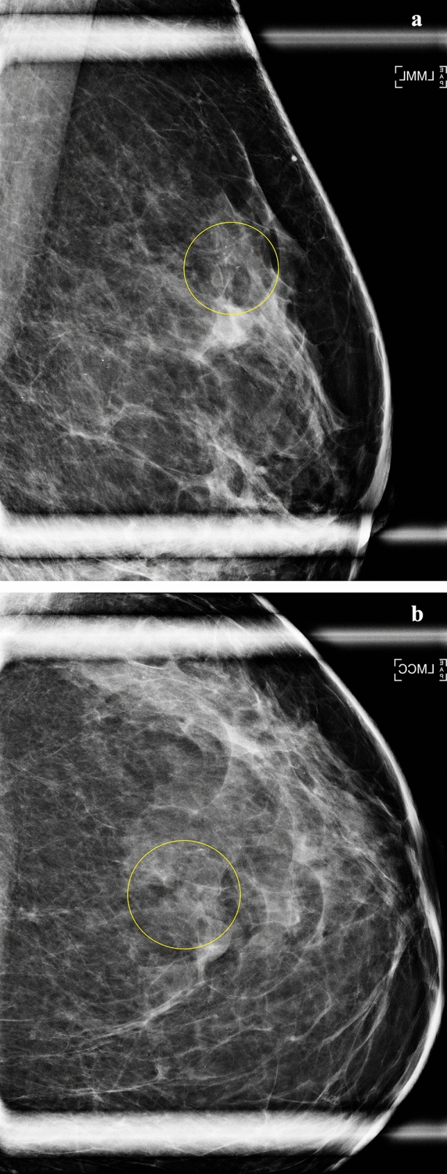
Purpose: Mammographic calcifications on mammograms with a negative/benign assessment are associated with increased breast cancer risk. Associations with advanced breast cancer risk are unknown. We evaluated whether calcifications recorded on mammography reports are associated with advanced invasive breast cancer risk.
Methods: We included 3,710,313 screening mammograms with a negative/benign final assessment performed on 991,991 women aged 40-74 in the Breast Cancer Surveillance Consortium associated with 7229 advanced cancers. We calculated cumulative 5-year advanced (prognostic pathologic stage ≥II) breast cancer risk and hazards ratios (HR) adjusted for clinical risk factors according to presence or absence of calcifications by menopausal status, dense (heterogeneously or extremely dense) vs. non-dense (almost entirely fatty or scattered fibroglandular density) breasts, body mass index (BMI) < 25 kg/m2 vs. ≥ 25 kg/m2.
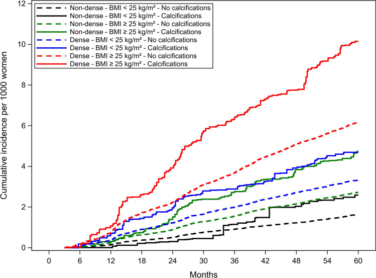
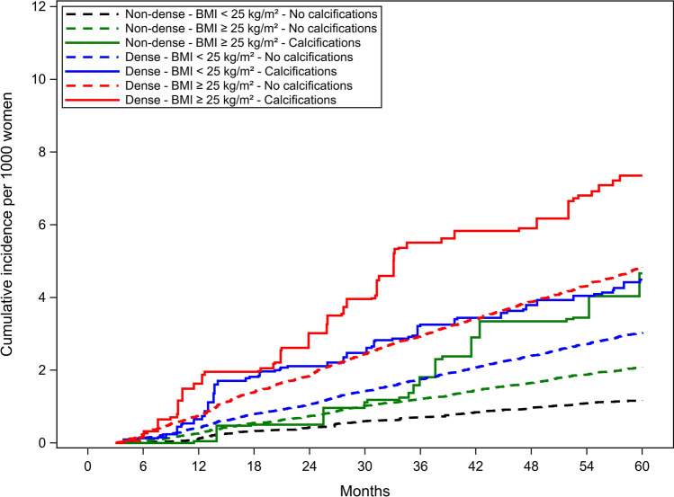
Results: Prevalence of calcifications was 6.1% among women who developed advanced breast cancer vs. 3.6% among others. Overall associations of advanced cancer with calcifications were similar for premenopausal (HR = 1.4; 95% CI 1.1-1.9) and postmenopausal (HR = 1.5; 95% CI 1.2-1.7) women. Compared to postmenopausal women with non-dense breasts and BMI < 25 kg/m2 without calcifications [cumulative 5-year advanced cancer incidence = 1.6 (95% CI 1.3-2.0) per 1000 women], postmenopausal women with dense breasts, BMI ≥ 25 kg/m2, and calcifications had 5.5-fold (95% CI 3.9-7.7) higher advanced cancer risk [cumulative 5-year advanced cancer incidence = 10.2; (95% CI 7.0-13.3) per 1000 women]. Results were similar for premenopausal women.
Conclusion: Mammographic calcifications increase advanced cancer risk beyond having dense breasts and being overweight/obese. Future research should investigate strength of associations by type of calcification and incorporation of calcifications into advanced cancer risk models for improvement in model accuracy.
目的:乳房x光检查中阴性/良性钙化与乳腺癌风险增加相关。与晚期乳腺癌风险的关系尚不清楚。我们评估了乳房x光检查报告中记录的钙化是否与晚期浸润性乳腺癌风险相关。方法:我们纳入了3710,313例最终评估为阴性/良性的筛查乳房x线照片,其中991,991例女性年龄在40-74岁之间,与7229例晚期癌症相关。我们计算了累积的5年晚期(预后病理分期≥II)乳腺癌风险和危险比(HR),根据绝经期是否存在钙化、乳房致密(非均匀或极致密)与非致密(几乎完全脂肪化或分散的纤维腺密度)、体重指数(BMI) 2与≥25 kg/m2调整了临床危险因素。结果:晚期乳腺癌女性的钙化患病率为6.1%,而其他女性为3.6%。绝经前晚期癌症与钙化的总体关联相似(HR = 1.4;95% CI 1.1-1.9)和绝经后(HR = 1.5;95% CI 1.2-1.7)。与绝经后乳房不致密且BMI 2无钙化的妇女相比[累计5年晚期癌症发病率= 1.6 (95% CI 1.3-2.0) / 1000名妇女],绝经后乳房致密且BMI≥25 kg/m2且钙化的妇女晚期癌症风险高5.5倍(95% CI 3.9-7.7)[累计5年晚期癌症发病率= 10.2;(95% CI 7.0-13.3)每1000名妇女]。绝经前妇女的结果相似。结论:乳房x线摄影钙化增加了晚期癌症的风险,超过了致密乳房和超重/肥胖。未来的研究应调查钙化类型的关联强度,并将钙化纳入晚期癌症风险模型,以提高模型的准确性。
期刊介绍:
Breast Cancer Research and Treatment provides the surgeon, radiotherapist, medical oncologist, endocrinologist, epidemiologist, immunologist or cell biologist investigating problems in breast cancer a single forum for communication. The journal creates a "market place" for breast cancer topics which cuts across all the usual lines of disciplines, providing a site for presenting pertinent investigations, and for discussing critical questions relevant to the entire field. It seeks to develop a new focus and new perspectives for all those concerned with breast cancer.
 求助内容:
求助内容: 应助结果提醒方式:


