Jie Lin, Hao Zheng, Yuan Dong, Lanqi Fu, Yujie Ding, Shucheng Huang, Shiwei Wang, Junna Wang
{"title":"Peritumoral Radiomic Features on CT for Differential Diagnosis in Small-Cell Lung Cancer: Potential for Surgical Decision-Making.","authors":"Jie Lin, Hao Zheng, Yuan Dong, Lanqi Fu, Yujie Ding, Shucheng Huang, Shiwei Wang, Junna Wang","doi":"10.1177/10732748251351754","DOIUrl":null,"url":null,"abstract":"<p><p><b>Introduction:</b> Small-cell lung cancer (SCLC) is a leading cause of cancer-related mortality worldwide, with limited therapeutic outcomes and poor prognosis. Accurate diagnosis and optimal surgical decision-making remain critical challenges. This study aimed to develop and validate a clinical-radiomics nomogram integrating computed tomography (CT) radiomic features of the peritumoral region and clinical factors to improve SCLC diagnosis and guide surgical planning.<b>Methods:</b> A retrospective cohort of 113 patients (54 SCLC, 59 non-small cell lung cancer) was analyzed. CT images were processed to extract 1050 radiomic features from both intratumoral and peritumoral (2-mm expanded) ROIs. Feature selection was performed using t-tests, LASSO regression, and mRMR analysis. Logistic regression models were constructed for original and expanded ROIs, and a clinical-radiomics nomogram was developed by combining significant radiomic features with independent clinical predictors (gender, smoking history, tumor diameter, glitch, and neuron-specific enolase levels). Model performance was evaluated using ROC curves, AUC, sensitivity, specificity, and CIC curves.<b>Results:</b> The expanded ROI radiomics model outperformed the original ROI and clinical models, achieving higher accuracy (0.83 vs 0.76/0.70), sensitivity (0.80 vs 0.74/0.77), specificity (0.85 vs 0.75/0.65), and AUC (0.85 vs 0.76/0.71). The clinical-radiomics nomogram demonstrated superior diagnostic performance, with an AUC of 0.96 (95% CI: 0.88-1.00), accuracy of 0.91, sensitivity of 0.92, and specificity of 0.90. CIC analysis confirmed its clinical utility for surgical decision-making at intermediate-risk thresholds.<b>Conclusion:</b> The integration of peritumoral radiomic features and clinical factors into a nomogram provides a non-invasive tool for SCLC diagnosis and surgical planning. The superiority of the expanded model substantiates the potential presence of SCLC in peri-tumoral tissues that may be imperceptible through conventional imaging, thereby offering guidance for surgical decision-making. This approach has potential for improving treatment outcomes and warrants further validation in multicenter studies.</p>","PeriodicalId":49093,"journal":{"name":"Cancer Control","volume":"32 ","pages":"10732748251351754"},"PeriodicalIF":2.6000,"publicationDate":"2025-01-01","publicationTypes":"Journal Article","fieldsOfStudy":null,"isOpenAccess":false,"openAccessPdf":"https://www.ncbi.nlm.nih.gov/pmc/articles/PMC12174677/pdf/","citationCount":"0","resultStr":null,"platform":"Semanticscholar","paperid":null,"PeriodicalName":"Cancer Control","FirstCategoryId":"3","ListUrlMain":"https://doi.org/10.1177/10732748251351754","RegionNum":4,"RegionCategory":"医学","ArticlePicture":[],"TitleCN":null,"AbstractTextCN":null,"PMCID":null,"EPubDate":"2025/6/16 0:00:00","PubModel":"Epub","JCR":"Q3","JCRName":"ONCOLOGY","Score":null,"Total":0}
引用次数: 0
Abstract
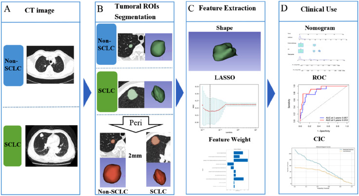
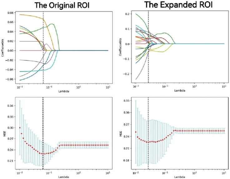
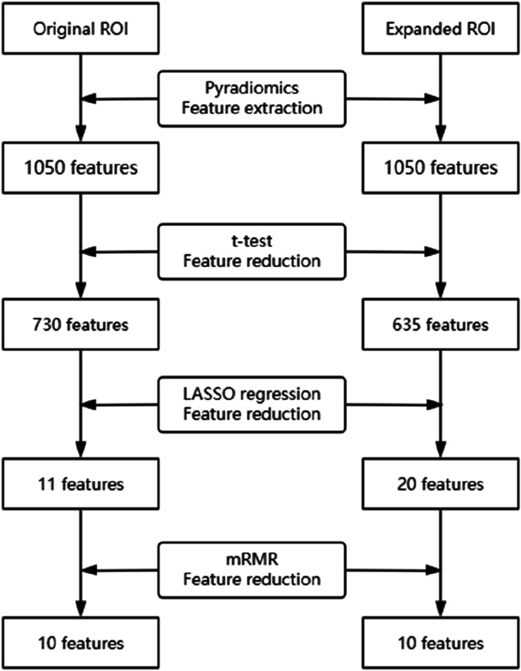
Introduction: Small-cell lung cancer (SCLC) is a leading cause of cancer-related mortality worldwide, with limited therapeutic outcomes and poor prognosis. Accurate diagnosis and optimal surgical decision-making remain critical challenges. This study aimed to develop and validate a clinical-radiomics nomogram integrating computed tomography (CT) radiomic features of the peritumoral region and clinical factors to improve SCLC diagnosis and guide surgical planning.Methods: A retrospective cohort of 113 patients (54 SCLC, 59 non-small cell lung cancer) was analyzed. CT images were processed to extract 1050 radiomic features from both intratumoral and peritumoral (2-mm expanded) ROIs. Feature selection was performed using t-tests, LASSO regression, and mRMR analysis. Logistic regression models were constructed for original and expanded ROIs, and a clinical-radiomics nomogram was developed by combining significant radiomic features with independent clinical predictors (gender, smoking history, tumor diameter, glitch, and neuron-specific enolase levels). Model performance was evaluated using ROC curves, AUC, sensitivity, specificity, and CIC curves.Results: The expanded ROI radiomics model outperformed the original ROI and clinical models, achieving higher accuracy (0.83 vs 0.76/0.70), sensitivity (0.80 vs 0.74/0.77), specificity (0.85 vs 0.75/0.65), and AUC (0.85 vs 0.76/0.71). The clinical-radiomics nomogram demonstrated superior diagnostic performance, with an AUC of 0.96 (95% CI: 0.88-1.00), accuracy of 0.91, sensitivity of 0.92, and specificity of 0.90. CIC analysis confirmed its clinical utility for surgical decision-making at intermediate-risk thresholds.Conclusion: The integration of peritumoral radiomic features and clinical factors into a nomogram provides a non-invasive tool for SCLC diagnosis and surgical planning. The superiority of the expanded model substantiates the potential presence of SCLC in peri-tumoral tissues that may be imperceptible through conventional imaging, thereby offering guidance for surgical decision-making. This approach has potential for improving treatment outcomes and warrants further validation in multicenter studies.
小细胞肺癌(SCLC)是全球癌症相关死亡的主要原因,治疗效果有限,预后差。准确的诊断和最佳的手术决策仍然是关键的挑战。本研究旨在开发和验证结合肿瘤周围区域CT放射学特征和临床因素的临床放射组学图,以提高SCLC的诊断和指导手术计划。方法:对113例患者(54例小细胞肺癌,59例非小细胞肺癌)进行回顾性分析。对CT图像进行处理,从瘤内和瘤周(2mm扩展)roi中提取1050个放射学特征。使用t检验、LASSO回归和mRMR分析进行特征选择。对原始roi和扩展roi构建了逻辑回归模型,并通过将显著的放射学特征与独立的临床预测因子(性别、吸烟史、肿瘤直径、glitch和神经元特异性烯醇化酶水平)相结合,建立了临床-放射组学nomogram。采用ROC曲线、AUC、敏感性、特异性和CIC曲线评价模型的性能。结果:扩展后的ROI放射组学模型优于原始ROI和临床模型,具有更高的准确性(0.83 vs 0.76/0.70)、灵敏度(0.80 vs 0.74/0.77)、特异性(0.85 vs 0.75/0.65)和AUC (0.85 vs 0.76/0.71)。临床放射组学影像学表现出优越的诊断性能,AUC为0.96 (95% CI: 0.88-1.00),准确性为0.91,敏感性为0.92,特异性为0.90。CIC分析证实了其在中等风险阈值下手术决策的临床应用。结论:将肿瘤周围放射学特征和临床因素整合到nomographic中,为SCLC的诊断和手术计划提供了一种无创工具。扩大模型的优越性证实了SCLC存在于肿瘤周围组织的可能性,这可能是通过常规成像无法察觉的,从而为手术决策提供指导。该方法具有改善治疗结果的潜力,值得在多中心研究中进一步验证。
期刊介绍:
Cancer Control is a JCR-ranked, peer-reviewed open access journal whose mission is to advance the prevention, detection, diagnosis, treatment, and palliative care of cancer by enabling researchers, doctors, policymakers, and other healthcare professionals to freely share research along the cancer control continuum. Our vision is a world where gold-standard cancer care is the norm, not the exception.
 求助内容:
求助内容: 应助结果提醒方式:


