{"title":"Axillary adipose tissue-derived lymphatic endothelial cells exhibit distinct transcriptomic signatures reflecting lymphatic invasion status in breast cancer.","authors":"Asumi Iesato, Jun Suzuka, Kazutaka Otsuji, Tomo Osako, Nami Yamashita, Yuka Inoue, Tetsuyo Maeda, Natsue Uehiro, Kazuyo Yoshida, Yoko Takahashi, Kohei Kumegawa, Sumito Saeki, Liying Yang, Ai Tsuchiya, Kana Sakiyama, Miwa Tanaka, Takehiko Sakai, Shinji Ohno, Tetsuo Noda, Takayuki Ueno, Reo Maruyama","doi":"10.1186/s13058-025-02067-w","DOIUrl":null,"url":null,"abstract":"<p><strong>Background: </strong>Lymphatics provide a route for breast cancer cells to metastasize. Lymphatic endothelial cells (LECs), which form the structure of lymphatic vessels, play a key role in this process. Although LECs are pivotal in cancer progression, studies often rely on commercially available cell lines that may not accurately reflect the tumor microenvironment. Therefore, there is a pressing need to directly study patient-derived LECs to better understand their role in breast cancer.</p><p><strong>Methods: </strong>This study developed a method to isolate and characterize LECs directly from human breast-to-axilla adipose tissue. We used magnetic cell separation to remove CD45 + leukocytes and fluorescence-activated cell sorting to isolate cells expressing CD31 and podoplanin. Isolated cells were cultured under conditions promoting endothelial cell growth and were characterized through various assays assessing proliferation, tube formation, and gene expression patterns.</p><p><strong>Results: </strong>The sorted CD31 + /PDPN + /CD45 - cell populations exhibited marked increases in proliferation upon VEGF-C stimulation and formed tubule structures on BME-coated dishes, confirming their LEC properties. Notably, isolated LECs showed distinct gene expression patterns depending on the presence of lymph node metastasis and lymphatic invasion.</p><p><strong>Conclusions: </strong>The ability to isolate and characterize patient-derived LECs from mammary adipose tissue offers new insights into the cellular mechanisms underlying breast cancer metastasis. Significant gene expression variability related to disease state highlights the potential of these cells as biomarkers and therapeutic targets. This study emphasizes the importance of using patient-derived cells to accurately assess the tumor microenvironment, potentially leading to more personalized therapeutic approaches.</p>","PeriodicalId":49227,"journal":{"name":"Breast Cancer Research","volume":"27 1","pages":"109"},"PeriodicalIF":5.6000,"publicationDate":"2025-06-17","publicationTypes":"Journal Article","fieldsOfStudy":null,"isOpenAccess":false,"openAccessPdf":"https://www.ncbi.nlm.nih.gov/pmc/articles/PMC12172209/pdf/","citationCount":"0","resultStr":null,"platform":"Semanticscholar","paperid":null,"PeriodicalName":"Breast Cancer Research","FirstCategoryId":"3","ListUrlMain":"https://doi.org/10.1186/s13058-025-02067-w","RegionNum":1,"RegionCategory":"医学","ArticlePicture":[],"TitleCN":null,"AbstractTextCN":null,"PMCID":null,"EPubDate":"","PubModel":"","JCR":"Q1","JCRName":"Medicine","Score":null,"Total":0}
引用次数: 0
Abstract
Background: Lymphatics provide a route for breast cancer cells to metastasize. Lymphatic endothelial cells (LECs), which form the structure of lymphatic vessels, play a key role in this process. Although LECs are pivotal in cancer progression, studies often rely on commercially available cell lines that may not accurately reflect the tumor microenvironment. Therefore, there is a pressing need to directly study patient-derived LECs to better understand their role in breast cancer.
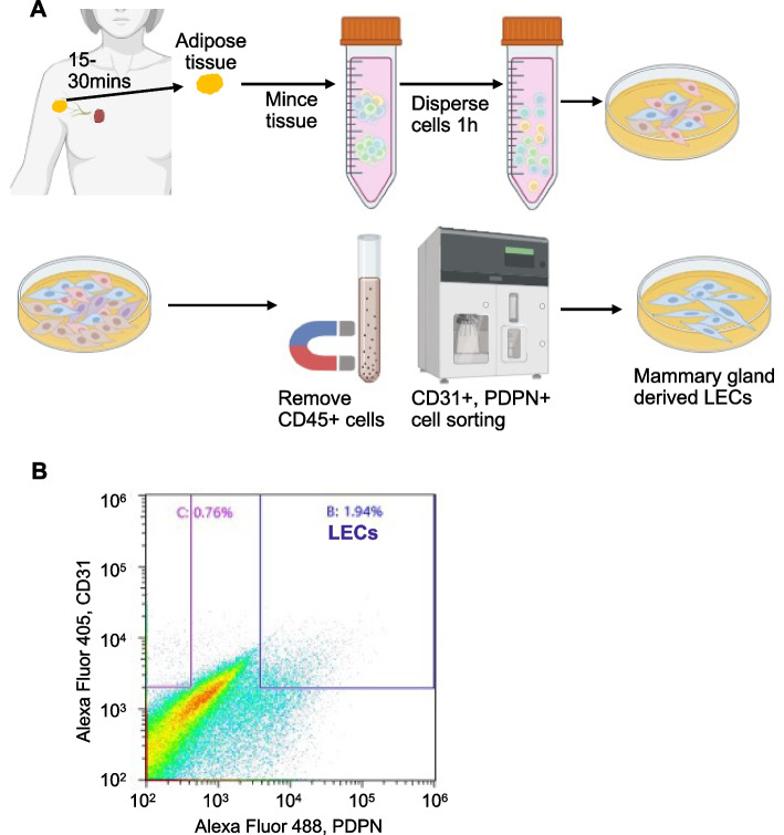
Methods: This study developed a method to isolate and characterize LECs directly from human breast-to-axilla adipose tissue. We used magnetic cell separation to remove CD45 + leukocytes and fluorescence-activated cell sorting to isolate cells expressing CD31 and podoplanin. Isolated cells were cultured under conditions promoting endothelial cell growth and were characterized through various assays assessing proliferation, tube formation, and gene expression patterns.
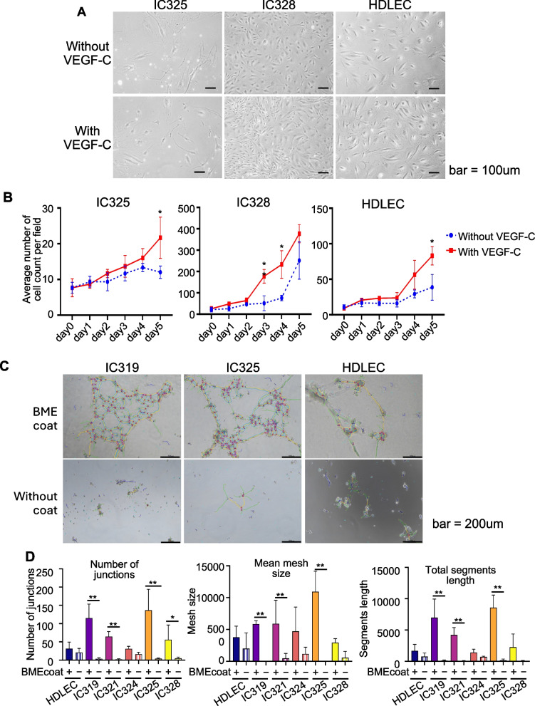
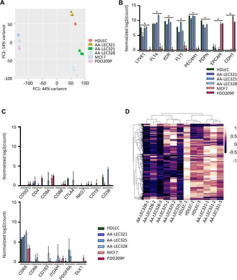
Results: The sorted CD31 + /PDPN + /CD45 - cell populations exhibited marked increases in proliferation upon VEGF-C stimulation and formed tubule structures on BME-coated dishes, confirming their LEC properties. Notably, isolated LECs showed distinct gene expression patterns depending on the presence of lymph node metastasis and lymphatic invasion.
Conclusions: The ability to isolate and characterize patient-derived LECs from mammary adipose tissue offers new insights into the cellular mechanisms underlying breast cancer metastasis. Significant gene expression variability related to disease state highlights the potential of these cells as biomarkers and therapeutic targets. This study emphasizes the importance of using patient-derived cells to accurately assess the tumor microenvironment, potentially leading to more personalized therapeutic approaches.
期刊介绍:
Breast Cancer Research, an international, peer-reviewed online journal, publishes original research, reviews, editorials, and reports. It features open-access research articles of exceptional interest across all areas of biology and medicine relevant to breast cancer. This includes normal mammary gland biology, with a special emphasis on the genetic, biochemical, and cellular basis of breast cancer. In addition to basic research, the journal covers preclinical, translational, and clinical studies with a biological basis, including Phase I and Phase II trials.

 求助内容:
求助内容: 应助结果提醒方式:


