Ali Naimi, Paul Martin Putora, Christian Rothermundt, Antonia Digklia, Jose Manuel Asencio, Sylvie Bonvalot, Florian Bösch, Anant Desai, Amer James Durrani, Haim Gutman, Daphne Hompes, Jens Jakob, Wolfram Trudo Knoefel, Elisabetta Pennacchioli, Piotr Rutkowski, Winan J van Houdt, Barbara L van Leeuwen, Stephan Waelti, Tim Steffen Fischer, Stefan Markart, Simon Wildermuth, Tobias Johannes Dietrich
{"title":"Diagnostic work-up of lipomatous tumors: a decision-making analysis among European sarcoma centers.","authors":"Ali Naimi, Paul Martin Putora, Christian Rothermundt, Antonia Digklia, Jose Manuel Asencio, Sylvie Bonvalot, Florian Bösch, Anant Desai, Amer James Durrani, Haim Gutman, Daphne Hompes, Jens Jakob, Wolfram Trudo Knoefel, Elisabetta Pennacchioli, Piotr Rutkowski, Winan J van Houdt, Barbara L van Leeuwen, Stephan Waelti, Tim Steffen Fischer, Stefan Markart, Simon Wildermuth, Tobias Johannes Dietrich","doi":"10.1186/s13244-025-02012-7","DOIUrl":null,"url":null,"abstract":"<p><strong>Objectives: </strong>Lipomatous soft-tissue tumors present a diagnostic burden. The aim of this work was to compare standard operating procedures (SOPs) for the diagnostic management of lipomatous soft-tissue tumors among European academic centers.</p><p><strong>Methods: </strong>Experts of the Soft Tissue and Bone Sarcoma Group of the European Organization for Research and Treatment of Cancer were asked for their SOPs in the diagnosis of adipocytic soft-tissue tumors in an otherwise healthy patient. The answers were converted to decision trees and subsequently compared using the objective consensus methodology. Mediastinal and retroperitoneal lipomatous tumors were excluded from the analysis.</p><p><strong>Results: </strong>The highest consensus (93%) among fourteen institutions was noted for evaluation with core needle biopsy (CNB) as SOP for lipomatous tumors located deep in tissue exceeding 7 cm and tumor-associated symptoms. Evaluation of heterogeneous features on imaging by CNB usually showed a consensus rate of at least 75%. Consensus was less likely for lipomatous tumors without symptoms or heterogeneous features. In these settings, CNB and follow-up were almost equally recommended. For lipomatous tumors smaller than 3 cm, without growth or symptoms, no localization in the trunk, and homogeneous imaging features, a consensus rate of 71% was achieved for follow-up.</p><p><strong>Conclusions: </strong>SOPs for diagnostic work-up of lipomatous tumors varied despite their geographical proximity. The highest consensus for biopsy was for deep large tumors with associated symptoms. For follow-up, consensus was shown for small homogenous tumors outside the trunk, without growth or symptoms. Consensus on resection involved homogeneous deeply located small tumors outside the trunk with growth and symptoms.</p><p><strong>Critical relevance statement: </strong>This study identifies the decision-making criteria with the highest consensus rate among participating academic sarcoma centers in diagnosing lipomatous tumors: tumors located deep in the tissue, a tumor size exceeding 7 cm, and associated symptoms emerge as pivotal criteria.</p><p><strong>Key points: </strong>Standard operating procedures for diagnostic work-up of lipomatous tumors among fourteen sarcoma centers were analyzed. Identified diagnostic criteria are: imaging features, size, growth, symptoms, superficial and trunk location. The highest consensus concerned recommending biopsies for deep tumors > 7 cm with associated symptoms.</p>","PeriodicalId":13639,"journal":{"name":"Insights into Imaging","volume":"16 1","pages":"123"},"PeriodicalIF":4.5000,"publicationDate":"2025-06-14","publicationTypes":"Journal Article","fieldsOfStudy":null,"isOpenAccess":false,"openAccessPdf":"https://www.ncbi.nlm.nih.gov/pmc/articles/PMC12167213/pdf/","citationCount":"0","resultStr":null,"platform":"Semanticscholar","paperid":null,"PeriodicalName":"Insights into Imaging","FirstCategoryId":"3","ListUrlMain":"https://doi.org/10.1186/s13244-025-02012-7","RegionNum":2,"RegionCategory":"医学","ArticlePicture":[],"TitleCN":null,"AbstractTextCN":null,"PMCID":null,"EPubDate":"","PubModel":"","JCR":"Q1","JCRName":"RADIOLOGY, NUCLEAR MEDICINE & MEDICAL IMAGING","Score":null,"Total":0}
引用次数: 0
Abstract
Objectives: Lipomatous soft-tissue tumors present a diagnostic burden. The aim of this work was to compare standard operating procedures (SOPs) for the diagnostic management of lipomatous soft-tissue tumors among European academic centers.
Methods: Experts of the Soft Tissue and Bone Sarcoma Group of the European Organization for Research and Treatment of Cancer were asked for their SOPs in the diagnosis of adipocytic soft-tissue tumors in an otherwise healthy patient. The answers were converted to decision trees and subsequently compared using the objective consensus methodology. Mediastinal and retroperitoneal lipomatous tumors were excluded from the analysis.
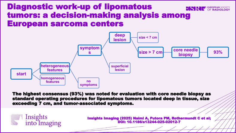
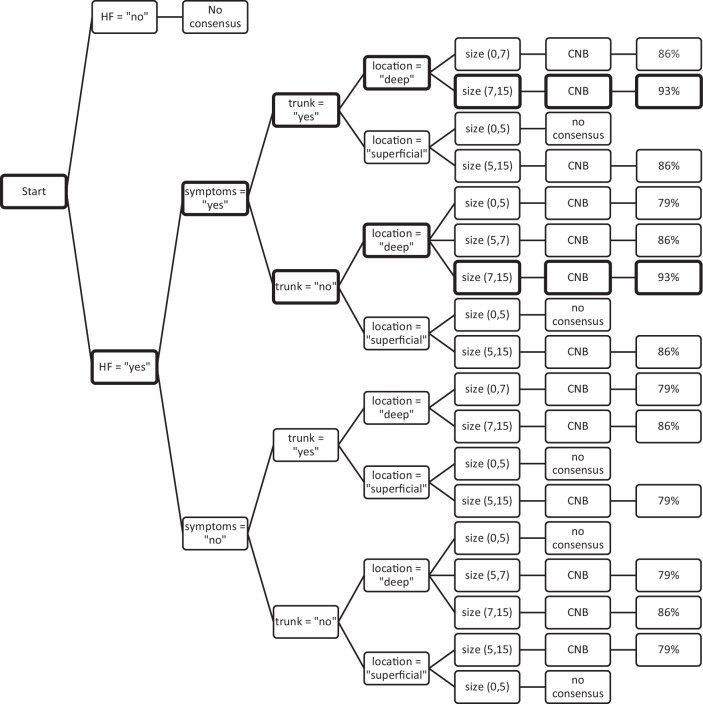
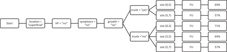
Results: The highest consensus (93%) among fourteen institutions was noted for evaluation with core needle biopsy (CNB) as SOP for lipomatous tumors located deep in tissue exceeding 7 cm and tumor-associated symptoms. Evaluation of heterogeneous features on imaging by CNB usually showed a consensus rate of at least 75%. Consensus was less likely for lipomatous tumors without symptoms or heterogeneous features. In these settings, CNB and follow-up were almost equally recommended. For lipomatous tumors smaller than 3 cm, without growth or symptoms, no localization in the trunk, and homogeneous imaging features, a consensus rate of 71% was achieved for follow-up.
Conclusions: SOPs for diagnostic work-up of lipomatous tumors varied despite their geographical proximity. The highest consensus for biopsy was for deep large tumors with associated symptoms. For follow-up, consensus was shown for small homogenous tumors outside the trunk, without growth or symptoms. Consensus on resection involved homogeneous deeply located small tumors outside the trunk with growth and symptoms.
Critical relevance statement: This study identifies the decision-making criteria with the highest consensus rate among participating academic sarcoma centers in diagnosing lipomatous tumors: tumors located deep in the tissue, a tumor size exceeding 7 cm, and associated symptoms emerge as pivotal criteria.
Key points: Standard operating procedures for diagnostic work-up of lipomatous tumors among fourteen sarcoma centers were analyzed. Identified diagnostic criteria are: imaging features, size, growth, symptoms, superficial and trunk location. The highest consensus concerned recommending biopsies for deep tumors > 7 cm with associated symptoms.
期刊介绍:
Insights into Imaging (I³) is a peer-reviewed open access journal published under the brand SpringerOpen. All content published in the journal is freely available online to anyone, anywhere!
I³ continuously updates scientific knowledge and progress in best-practice standards in radiology through the publication of original articles and state-of-the-art reviews and opinions, along with recommendations and statements from the leading radiological societies in Europe.
Founded by the European Society of Radiology (ESR), I³ creates a platform for educational material, guidelines and recommendations, and a forum for topics of controversy.
A balanced combination of review articles, original papers, short communications from European radiological congresses and information on society matters makes I³ an indispensable source for current information in this field.
I³ is owned by the ESR, however authors retain copyright to their article according to the Creative Commons Attribution License (see Copyright and License Agreement). All articles can be read, redistributed and reused for free, as long as the author of the original work is cited properly.
The open access fees (article-processing charges) for this journal are kindly sponsored by ESR for all Members.
The journal went open access in 2012, which means that all articles published since then are freely available online.

 求助内容:
求助内容: 应助结果提醒方式:


