Federica Pediconi, Annarita Speranza, Giuliana Moffa, Roberto Maroncelli, Sara Coppola, Francesca Galati, Claudia Bernardi, Giacomo Maccagno, Dominga Pugliese, Carlo Catalano, Andrea Laghi, Veronica Rizzo
{"title":"Contrast-enhanced mammography for breast cancer detection and diagnosis with high concentration iodinated contrast medium.","authors":"Federica Pediconi, Annarita Speranza, Giuliana Moffa, Roberto Maroncelli, Sara Coppola, Francesca Galati, Claudia Bernardi, Giacomo Maccagno, Dominga Pugliese, Carlo Catalano, Andrea Laghi, Veronica Rizzo","doi":"10.1186/s13244-025-01994-8","DOIUrl":null,"url":null,"abstract":"<p><strong>Objectives: </strong>We assessed the diagnostic performance of contrast-enhanced mammography (CEM) using a high-concentration iodinated contrast medium (HCCM, 400 mgI/mL) to determine whether the reduced iodine dose and increased iodine delivery rate (IDR) achieved might offer a more sustainable alternative to CEM performed with lower iodine concentrations.</p><p><strong>Methods: </strong>This two-center retrospective study included 205 patients who underwent CEM between March 2021 and February 2022. Patients were injected with HCCM at 1.0 mL/kg bodyweight at an IDR of 1.2 gL/s. Standard cranio-caudal and mediolateral-oblique views were acquired. Images were reviewed independently by two experienced radiologists who were blinded to patient clinical and imaging information. Diagnostic performance, including sensitivity, specificity, and accuracy, was assessed based on histological or long-term imaging follow-up as the reference standard.</p><p><strong>Results: </strong>Among the 205 patients, 149 (72.7%) had malignant lesions, and 56 (27.3%) had benign findings. The sensitivity and specificity of CEM were 96-97% and 84-87.5%, respectively, with an overall accuracy of 93-95%. The IDR achieved with HCCM resulted in excellent contrast enhancement, particularly in patients with aggressive malignancies. ROC analysis confirmed the good diagnostic performance, with AUC values of 0.90-0.92. Compared to conventional mammography and ultrasound, CEM demonstrated significantly higher diagnostic accuracy, especially in patients with dense breast tissue.</p><p><strong>Conclusions: </strong>CEM with HCCM provides excellent diagnostic performance, achieving high sensitivity and specificity while allowing for a reduced iodine dose and increased IDR. This approach may offer a more sustainable alternative to conventional contrast media without compromising diagnostic accuracy, particularly for the detection and characterization of aggressive breast lesions.</p><p><strong>Critical relevance statement: </strong>This study demonstrates that reducing the volume of injected contrast media while increasing iodine concentration maintains the diagnostic benefits of CEM, further supporting its potential to improve early cancer detection, thereby advancing clinical radiology practices and optimizing screening strategies for women with dense breasts.</p><p><strong>Key points: </strong>Currently, CEM protocols utilize a variety of iodine concentrations and flow rates. CEM with high-concentration contrast (400 mgI/mL) achieved 96% sensitivity and 87.5% specificity. High-concentration contrast in CEM improves early detection of aggressive breast cancers.</p>","PeriodicalId":13639,"journal":{"name":"Insights into Imaging","volume":"16 1","pages":"124"},"PeriodicalIF":4.5000,"publicationDate":"2025-06-14","publicationTypes":"Journal Article","fieldsOfStudy":null,"isOpenAccess":false,"openAccessPdf":"https://www.ncbi.nlm.nih.gov/pmc/articles/PMC12167179/pdf/","citationCount":"0","resultStr":null,"platform":"Semanticscholar","paperid":null,"PeriodicalName":"Insights into Imaging","FirstCategoryId":"3","ListUrlMain":"https://doi.org/10.1186/s13244-025-01994-8","RegionNum":2,"RegionCategory":"医学","ArticlePicture":[],"TitleCN":null,"AbstractTextCN":null,"PMCID":null,"EPubDate":"","PubModel":"","JCR":"Q1","JCRName":"RADIOLOGY, NUCLEAR MEDICINE & MEDICAL IMAGING","Score":null,"Total":0}
引用次数: 0
Abstract
Objectives: We assessed the diagnostic performance of contrast-enhanced mammography (CEM) using a high-concentration iodinated contrast medium (HCCM, 400 mgI/mL) to determine whether the reduced iodine dose and increased iodine delivery rate (IDR) achieved might offer a more sustainable alternative to CEM performed with lower iodine concentrations.
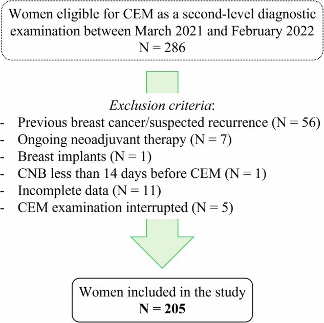
Methods: This two-center retrospective study included 205 patients who underwent CEM between March 2021 and February 2022. Patients were injected with HCCM at 1.0 mL/kg bodyweight at an IDR of 1.2 gL/s. Standard cranio-caudal and mediolateral-oblique views were acquired. Images were reviewed independently by two experienced radiologists who were blinded to patient clinical and imaging information. Diagnostic performance, including sensitivity, specificity, and accuracy, was assessed based on histological or long-term imaging follow-up as the reference standard.
Results: Among the 205 patients, 149 (72.7%) had malignant lesions, and 56 (27.3%) had benign findings. The sensitivity and specificity of CEM were 96-97% and 84-87.5%, respectively, with an overall accuracy of 93-95%. The IDR achieved with HCCM resulted in excellent contrast enhancement, particularly in patients with aggressive malignancies. ROC analysis confirmed the good diagnostic performance, with AUC values of 0.90-0.92. Compared to conventional mammography and ultrasound, CEM demonstrated significantly higher diagnostic accuracy, especially in patients with dense breast tissue.
Conclusions: CEM with HCCM provides excellent diagnostic performance, achieving high sensitivity and specificity while allowing for a reduced iodine dose and increased IDR. This approach may offer a more sustainable alternative to conventional contrast media without compromising diagnostic accuracy, particularly for the detection and characterization of aggressive breast lesions.
Critical relevance statement: This study demonstrates that reducing the volume of injected contrast media while increasing iodine concentration maintains the diagnostic benefits of CEM, further supporting its potential to improve early cancer detection, thereby advancing clinical radiology practices and optimizing screening strategies for women with dense breasts.
Key points: Currently, CEM protocols utilize a variety of iodine concentrations and flow rates. CEM with high-concentration contrast (400 mgI/mL) achieved 96% sensitivity and 87.5% specificity. High-concentration contrast in CEM improves early detection of aggressive breast cancers.
期刊介绍:
Insights into Imaging (I³) is a peer-reviewed open access journal published under the brand SpringerOpen. All content published in the journal is freely available online to anyone, anywhere!
I³ continuously updates scientific knowledge and progress in best-practice standards in radiology through the publication of original articles and state-of-the-art reviews and opinions, along with recommendations and statements from the leading radiological societies in Europe.
Founded by the European Society of Radiology (ESR), I³ creates a platform for educational material, guidelines and recommendations, and a forum for topics of controversy.
A balanced combination of review articles, original papers, short communications from European radiological congresses and information on society matters makes I³ an indispensable source for current information in this field.
I³ is owned by the ESR, however authors retain copyright to their article according to the Creative Commons Attribution License (see Copyright and License Agreement). All articles can be read, redistributed and reused for free, as long as the author of the original work is cited properly.
The open access fees (article-processing charges) for this journal are kindly sponsored by ESR for all Members.
The journal went open access in 2012, which means that all articles published since then are freely available online.

 求助内容:
求助内容: 应助结果提醒方式:


