Matthew J Fata, Steven M Hollenberg, Brent Klinkhammer, David Landers, George Rockett, Jana Tancredi, Zoltan Turi, Joseph E Parrillo
{"title":"Reversible Myocardial Depression and Dilatation in COVID-19 Shock Patients.","authors":"Matthew J Fata, Steven M Hollenberg, Brent Klinkhammer, David Landers, George Rockett, Jana Tancredi, Zoltan Turi, Joseph E Parrillo","doi":"10.1097/CCE.0000000000001264","DOIUrl":null,"url":null,"abstract":"<p><strong>Importance: </strong>A characteristic pattern of bacterial and fungal septic shock is decreased left ventricular (LV) ejection fraction (LVEF) and modest dilatation of the LV. In survivors, the myocardial depression and dilatation are reversible within several days. In a cohort of 368 hospitalized COVID patients with shock from March 2020 to December 2021, 15 patients were identified with an echocardiogram determined depressed LVEF during acute shock, and a follow-up echocardiogram was performed.</p><p><strong>Objectives: </strong>Myocardial dysfunction and dilatation associated with COVID-19 are reversible.</p><p><strong>Design, setting, and participants: </strong>LVEF was determined by Simpson's rule and stroke volume (SV) was analyzed by Doppler. Based on the LVEF and cardiac index (CI), patients were categorized into groups with low or normal values using an ejection fraction of 45% and CI 2.2 L/min/m2 as the respective thresholds. A subset of 15 patients underwent serial echocardiography, which was performed at a median of 13 days (95% CI, 9-39 d) after the initial value.</p><p><strong>Main outcomes and measures: </strong>The LVEF and LV volumes recorded during initial and follow-up echo were analyzed using paired t test.</p><p><strong>Results: </strong>Comparing initial during acute shock with follow-up values, the mean (± sd) LVEF was 35.3 ± 8.1 vs. 43.8 ± 3.47 (p = 0.031), indexed SV 29.6 ± 1.9 mL vs. 31.7 ± 2.3 mL (p = 0.522), LV end-diastolic volume 182 ± 14.1 mL vs. 152.1 ± 12.9 mL (p = 0.025), and LV end-systolic volume 120.2 ± 13.1 mL vs. 90.1 ± 12.1 mL (p = 0.025), respectively.</p><p><strong>Conclusions and relevance: </strong>Serial echocardiographic studies of COVID-19 shock patients with reduced LVEF and ventricular dilatation demonstrate reversibility of myocardial depression and dilation with no change in SV, a finding strikingly similar to that seen in bacterial and fungal-induced septic shock. Thus, COVID-19 (viral) induced septic shock may have a similar pathogenetic mechanism of myocardial dysfunction to that seen with bacterial or fungal sepsis.</p>","PeriodicalId":93957,"journal":{"name":"Critical care explorations","volume":"7 6","pages":"e1264"},"PeriodicalIF":2.7000,"publicationDate":"2025-06-13","publicationTypes":"Journal Article","fieldsOfStudy":null,"isOpenAccess":false,"openAccessPdf":"https://www.ncbi.nlm.nih.gov/pmc/articles/PMC12169969/pdf/","citationCount":"0","resultStr":null,"platform":"Semanticscholar","paperid":null,"PeriodicalName":"Critical care explorations","FirstCategoryId":"1085","ListUrlMain":"https://doi.org/10.1097/CCE.0000000000001264","RegionNum":0,"RegionCategory":null,"ArticlePicture":[],"TitleCN":null,"AbstractTextCN":null,"PMCID":null,"EPubDate":"2025/6/1 0:00:00","PubModel":"eCollection","JCR":"Q4","JCRName":"Medicine","Score":null,"Total":0}
引用次数: 0
Abstract
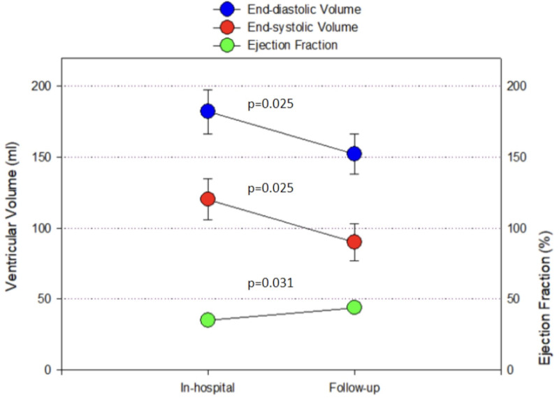
Importance: A characteristic pattern of bacterial and fungal septic shock is decreased left ventricular (LV) ejection fraction (LVEF) and modest dilatation of the LV. In survivors, the myocardial depression and dilatation are reversible within several days. In a cohort of 368 hospitalized COVID patients with shock from March 2020 to December 2021, 15 patients were identified with an echocardiogram determined depressed LVEF during acute shock, and a follow-up echocardiogram was performed.
Objectives: Myocardial dysfunction and dilatation associated with COVID-19 are reversible.
Design, setting, and participants: LVEF was determined by Simpson's rule and stroke volume (SV) was analyzed by Doppler. Based on the LVEF and cardiac index (CI), patients were categorized into groups with low or normal values using an ejection fraction of 45% and CI 2.2 L/min/m2 as the respective thresholds. A subset of 15 patients underwent serial echocardiography, which was performed at a median of 13 days (95% CI, 9-39 d) after the initial value.
Main outcomes and measures: The LVEF and LV volumes recorded during initial and follow-up echo were analyzed using paired t test.
Results: Comparing initial during acute shock with follow-up values, the mean (± sd) LVEF was 35.3 ± 8.1 vs. 43.8 ± 3.47 (p = 0.031), indexed SV 29.6 ± 1.9 mL vs. 31.7 ± 2.3 mL (p = 0.522), LV end-diastolic volume 182 ± 14.1 mL vs. 152.1 ± 12.9 mL (p = 0.025), and LV end-systolic volume 120.2 ± 13.1 mL vs. 90.1 ± 12.1 mL (p = 0.025), respectively.
Conclusions and relevance: Serial echocardiographic studies of COVID-19 shock patients with reduced LVEF and ventricular dilatation demonstrate reversibility of myocardial depression and dilation with no change in SV, a finding strikingly similar to that seen in bacterial and fungal-induced septic shock. Thus, COVID-19 (viral) induced septic shock may have a similar pathogenetic mechanism of myocardial dysfunction to that seen with bacterial or fungal sepsis.
重要性:细菌性和真菌性感染性休克的特征是左室射血分数(LVEF)降低和左室适度扩张。在幸存者中,心肌收缩和扩张在几天内是可逆的。在2020年3月至2021年12月期间住院的368例新冠肺炎休克患者中,15例患者在急性休克期间被超声心动图确定为LVEF下降,并进行了随访超声心动图检查。目的:与COVID-19相关的心肌功能障碍和扩张是可逆的。设计、环境和参与者:LVEF采用辛普森规则测定,卒中容积(SV)采用多普勒分析。根据LVEF和心脏指数(CI),分别以射血分数45%和CI 2.2 L/min/m2为阈值,将患者分为低或正常值组。15名患者接受了连续超声心动图检查,在初始值后的中位时间为13天(95% CI, 9-39 d)。主要观察指标:采用配对t检验分析初始和随访回声记录的LVEF和LV容积。结果:急性休克初期与随访值比较,平均(±sd) LVEF为35.3±8.1 vs. 43.8±3.47 (p = 0.031),指标SV为29.6±1.9 mL vs. 31.7±2.3 mL (p = 0.522),左室舒张末期容积为182±14.1 mL vs. 152.1±12.9 mL (p = 0.025),左室收缩末期容积为120.2±13.1 mL vs. 90.1±12.1 mL (p = 0.025)。结论及相关性:对LVEF降低和心室扩张的COVID-19休克患者进行的一系列超声心动图研究表明,心肌抑制和扩张是可逆性的,SV没有变化,这一发现与细菌和真菌引起的感染性休克非常相似。因此,COVID-19(病毒)诱导的感染性休克可能具有与细菌性或真菌性败血症相似的心肌功能障碍发病机制。

 求助内容:
求助内容: 应助结果提醒方式:


